Kun Tomasz, Jakubowski Lucjusz
Department of General and Experimental Pathology, Medical University in Łódź, Łódź, Poland.
Pol J Radiol. 2012 Jul;77(3):19-24. doi: 10.12659/pjr.883370.
Mast cells, owing to diversity of secreted mediators, play a crucial role in the regulation of inflammatory response. Together with basophils, mast cells constitute a central pathogenetic element of anaphylactic (IgE-dependent) and anaphylactoid (IgE-independent) reactions. In severe cases, generalized degranulation of mast cells may cause symptoms of anaphylactic shock. The influence of the classical, iodine-based contrast media on mastocyte degranulation has been fully described. Our objective was to determine the influence of the gadolinium-based MRI contrast media on histamine release from mast cells and to compare the activity of ionic and non-ionic preparations of contrast media.
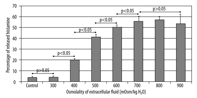
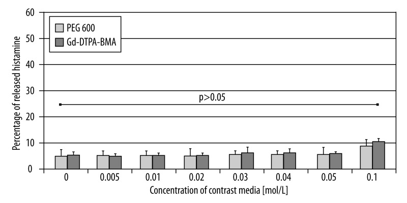
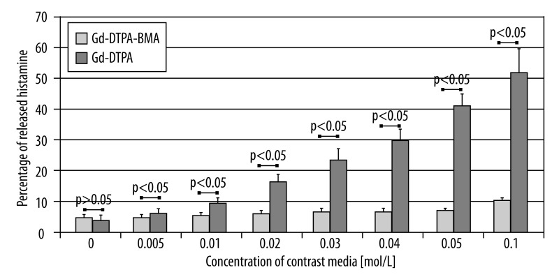
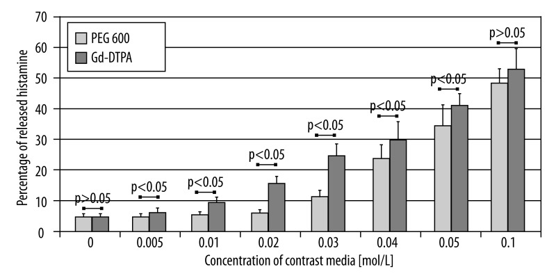
MATERIAL/METHODS: To determine the intensity of mast cell degranulation, we used an experimental model based on mastocytes isolated from rat peritoneal fluid. Purified suspensions of mast cells were incubated with various concentrations of Gd-DTPA and Gd-DTPA-BMA, and solutions of PEG 600 which served as a non-toxic osmotic stimulus. The intensity of mast cell activation was presented as mean percentage of histamine released from cells after incubation.
RESULTS/CONCLUSIONS: The obtained results demonstrate that both ionic and non-ionic preparations of the MRI contrast media are able to induce mast cell degranulation in vitro. It was also proved that the non-ionic MRI contrast media stimulate mast cells markedly more weakly than ionic contrast media at identical concentration. The aforementioned results may suggest a more profitable safety profile of the non-ionic contrast preparations. We may also conclude that triggering of mast cell degranulation after incubation with the solutions of MRI contrast media results from non-specific osmotic stimulation and direct toxicity of free ionic residues.
肥大细胞由于分泌介质的多样性,在炎症反应调节中起关键作用。肥大细胞与嗜碱性粒细胞一起,构成过敏(IgE 依赖性)和类过敏(IgE 非依赖性)反应的核心致病因素。在严重情况下,肥大细胞的全身性脱颗粒可能导致过敏性休克症状。基于碘的传统造影剂对肥大细胞脱颗粒的影响已得到充分描述。我们的目的是确定钆基 MRI 造影剂对肥大细胞组胺释放的影响,并比较造影剂离子型和非离子型制剂的活性。
材料/方法:为了确定肥大细胞脱颗粒的强度,我们使用了基于从大鼠腹腔液中分离出的肥大细胞的实验模型。将纯化的肥大细胞悬液与不同浓度的钆喷酸葡胺(Gd-DTPA)、钆布醇(Gd-DTPA-BMA)以及用作无毒渗透刺激物的聚乙二醇 600(PEG 600)溶液一起孵育。肥大细胞活化强度以孵育后细胞释放组胺的平均百分比表示。
结果/结论:所得结果表明,MRI 造影剂的离子型和非离子型制剂在体外均能诱导肥大细胞脱颗粒。还证明了在相同浓度下,非离子型 MRI 造影剂刺激肥大细胞的能力明显弱于离子型造影剂。上述结果可能表明非离子型造影剂制剂具有更有利的安全性。我们还可以得出结论,与 MRI 造影剂溶液孵育后肥大细胞脱颗粒的触发是由非特异性渗透刺激和游离离子残基的直接毒性引起的。