Kalogeropoulos Chris, Koumpoulis Ioannis, Papadiotis Evangelos, Zioga Aikaterini, Gkrepi Konstantina, Pappa Chrisavgi, Paschides Constantinos, Malamou-Mitsi Vasiliki, Aspiotis Miltiadis
Department of Ophthalmology, Medical School, University of Ioannina, Greece.
Clin Ophthalmol. 2012;6:1553-61. doi: 10.2147/OPTH.S34999. Epub 2012 Sep 24.
We describe two patients with squamous cell papilloma of the conjunctiva due to human papilloma virus (HPV) and review the literature.
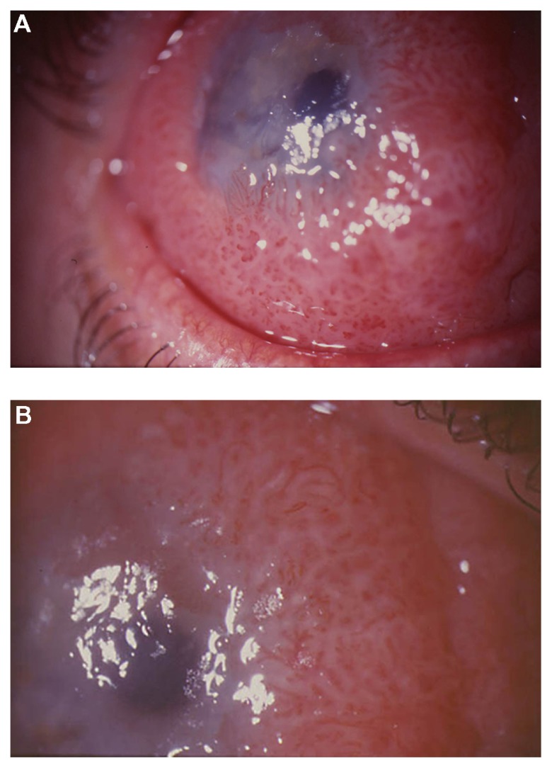
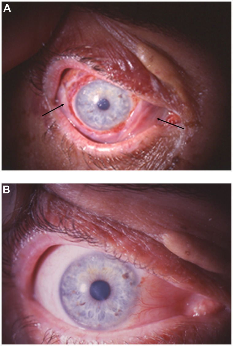
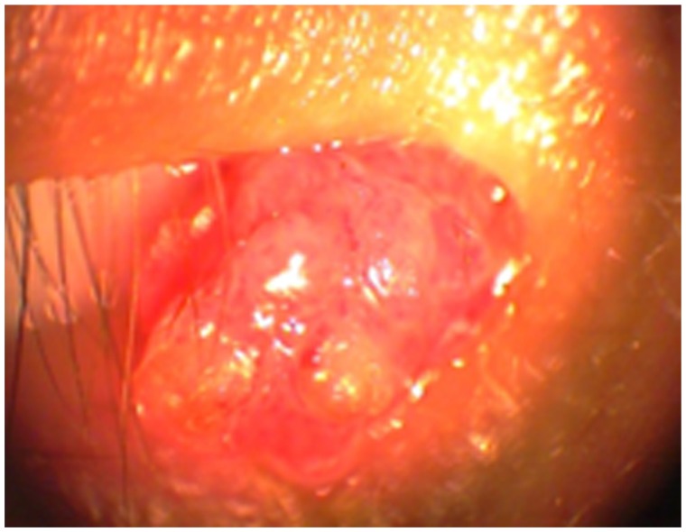
Two patients with conjunctival tumors were examined and treated in the University Eye Clinic and diagnosed in the University Pathology Department, University Hospital of Ioannina, Greece. The first patient was a 48-year-old man presenting with an extended papillomatous lesion in bulbar conjunctiva covering part of the cornea of his right eye. The second patient was a 24-year-old man presenting with a polypoidal papillomatous lesion on the caruncle of his right eye. The two lesions were removed surgically, cryotherapy was applied to the adjacent conjunctiva, and topical mitomycin-C was used. The amniotic membrane was used to restore the conjunctival defect in the first patient. The two removed lesions were sent to the Pathology Department for histopathological examination. Immunohistochemistry, DNA in situ hybridization, and polymerase chain reaction (PCR) analysis were performed.
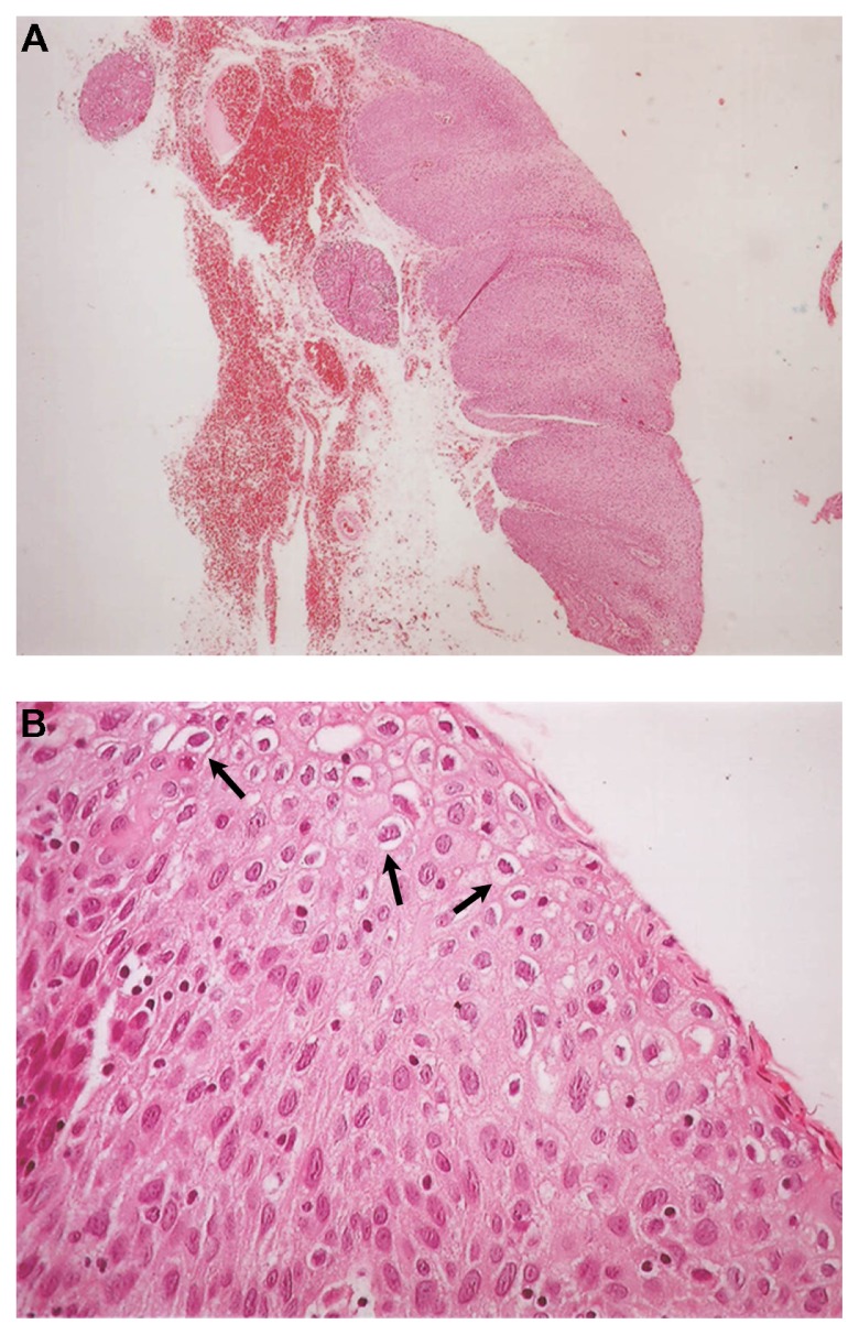
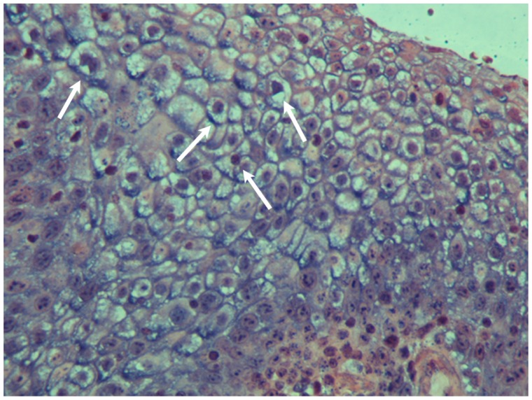
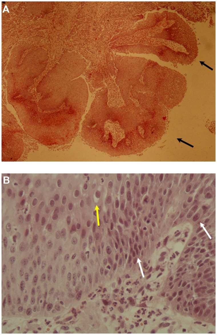
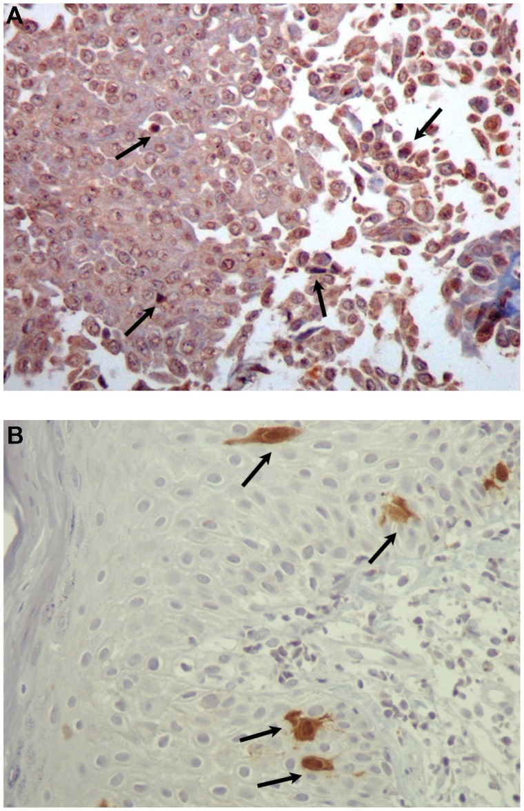
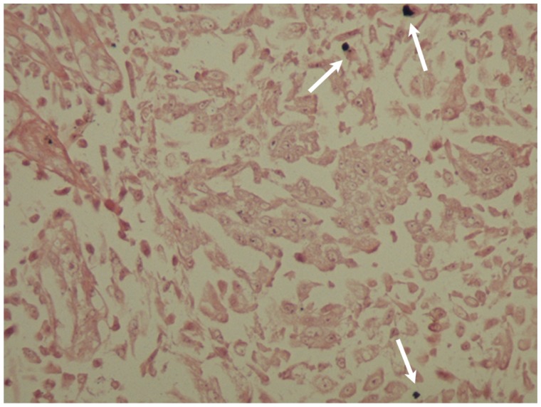
In the first patient, histopathology showed the presence of a benign squamous papilloma with koilocytosis. DNA in situ hybridization with broad-spectrum probes showed that this patient was positive for HPV DNA. In the second patient, histopathology showed the presence of a squamous papilloma with mild dysplasia and koilocytosis. Immunohistochemical analysis was positive for HPV protein and p16 protein. DNA in situ hybridization with broad-spectrum probes showed that the patient was positive for HPV DNA. PCR analysis showed the presence of HPV 6. According to morphological and molecular findings, both patients were diagnosed with squamous cell papilloma due to HPV.
HPV can infect the ocular surface. According to clinical results, the ophthalmologist in cooperation with the pathologist can recommend appropriate laboratory examinations to confirm the diagnosis and successfully treat conjunctival papillomas.
我们描述了两名因人类乳头瘤病毒(HPV)引起的结膜鳞状细胞乳头瘤患者,并对相关文献进行了综述。
两名结膜肿瘤患者在希腊约阿尼纳大学医院眼科诊所接受检查和治疗,并在该大学病理科进行诊断。第一名患者是一名48岁男性,右眼球结膜出现一个扩展的乳头瘤样病变,覆盖部分角膜。第二名患者是一名24岁男性,右眼泪阜出现一个息肉样乳头瘤样病变。两个病变均通过手术切除,对相邻结膜进行冷冻治疗,并使用局部丝裂霉素C。第一名患者使用羊膜修复结膜缺损。切除的两个病变送至病理科进行组织病理学检查。进行了免疫组织化学、DNA原位杂交和聚合酶链反应(PCR)分析。
在第一名患者中,组织病理学显示存在伴有空泡细胞的良性鳞状乳头瘤。使用广谱探针进行的DNA原位杂交显示该患者HPV DNA呈阳性。在第二名患者中,组织病理学显示存在伴有轻度发育异常和空泡细胞的鳞状乳头瘤。免疫组织化学分析显示HPV蛋白和p16蛋白呈阳性。使用广谱探针进行的DNA原位杂交显示该患者HPV DNA呈阳性。PCR分析显示存在HPV 6。根据形态学和分子学结果,两名患者均被诊断为因HPV引起的鳞状细胞乳头瘤。
HPV可感染眼表。根据临床结果,眼科医生与病理学家合作可推荐适当的实验室检查以确诊并成功治疗结膜乳头瘤。