Department of Pathology and Laboratory Medicine, University of Rochester Medical Center, 601 Elmwood Ave, Rochester, NY 14642, USA.
BMC Gastroenterol. 2012 Oct 18;12:146. doi: 10.1186/1471-230X-12-146.
High expression of Bmi-1, a key regulatory component of the polycomb repressive complex-1, has been associated with many solid and hematologic malignancies including esophageal squamous cell carcinoma. However, little is known about the role of Bmi-1 in esophageal adenocarcinoma. The aim of this study is to investigate the amplification and high expression of Bmi-1 and the associated clinicopathologic characteristics in esophageal adenocarcinoma and squamous cell carcinoma.
The protein expression level of Bmi-1 was detected by immunohistochemistry (IHC) from tissue microarrays (TMA) constructed at the University of Rochester from using tissues accrued between 1997 and 2005. Types of tissues included adenocarcinoma, squamous cell carcinoma and precancerous lesions. Patients' survival data, demographics, histologic diagnoses and tumor staging data were collected. The intensity (0-3) and percentage of Bmi-1 expression on TMA slides were scored by two pathologists. Genomic DNA from 116 esophageal adenocarcinoma was analyzed for copy number aberrations using Affymetrix SNP 6.0 arrays. Fisher exact tests and Kaplan-Meier methods were used to analyze data.
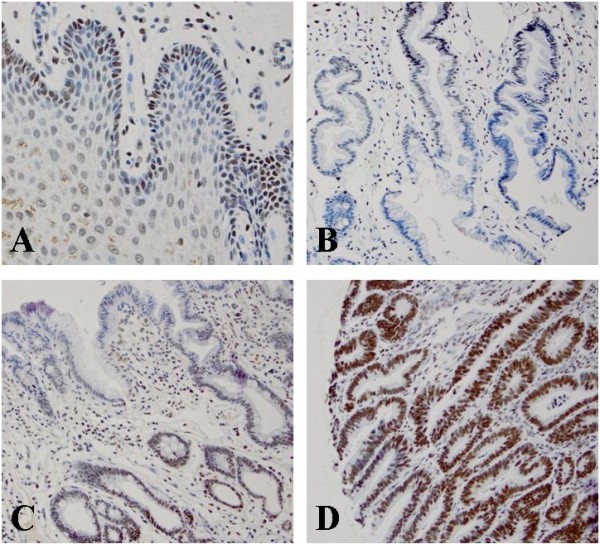
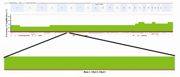
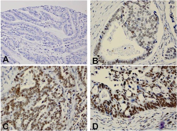
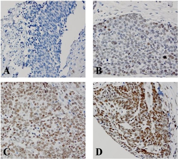
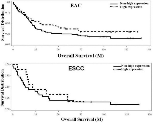
By IHC, Bmi-1 was focally expressed in the basal layers of almost all esophageal squamous mucosa, which was similar to previous reports in other organs related to stem cells. High Bmi-1 expression significantly increased from squamous epithelium (7%), columnar cell metaplasia (22%), Barrett's esophagus (22%), to low- (45%) and high-grade dysplasia (43%) and adenocarcinoma (37%). The expression level of Bmi-1 was significantly associated with esophageal adenocarcinoma differentiation. In esophageal adenocarcinoma, Bmi-1 amplification was detected by DNA microarray in a low percentage (3%). However, high Bmi-1 expression did not show an association with overall survival in both esophageal adenocarcinoma and squamous cell carcinoma.
This study demonstrates that high expression Bmi-1 is associated with esophageal adenocarcinoma and precancerous lesions, which implies that Bmi-1 plays an important role in early carcinogenesis in esophageal adenocarcinoma.
Bmi-1 是多梳抑制复合物-1 的关键调节成分,其高表达与包括食管鳞状细胞癌在内的许多实体瘤和血液恶性肿瘤有关。然而,关于 Bmi-1 在食管腺癌中的作用知之甚少。本研究旨在探讨 Bmi-1 的扩增和高表达及其与食管腺癌和鳞状细胞癌的临床病理特征的关系。
利用罗切斯特大学于 1997 年至 2005 年间收集的组织构建的组织微阵列(TMA),通过免疫组织化学(IHC)检测 Bmi-1 的蛋白表达水平。组织类型包括腺癌、鳞状细胞癌和癌前病变。收集了患者的生存数据、人口统计学数据、组织学诊断和肿瘤分期数据。两位病理学家对 TMA 切片上 Bmi-1 的表达强度(0-3)和百分比进行了评分。对 116 例食管腺癌的基因组 DNA 进行了 Affymetrix SNP 6.0 芯片分析,以检测拷贝数异常。采用 Fisher 确切检验和 Kaplan-Meier 方法进行数据分析。
通过 IHC,Bmi-1 在几乎所有食管鳞状黏膜的基底细胞层呈局灶性表达,这与其他与干细胞相关的器官的先前报道相似。高 Bmi-1 表达率从鳞状上皮(7%)、柱状细胞化生(22%)、Barrett 食管(22%)逐渐增加到低级别(45%)和高级别(43%)异型增生和腺癌(37%)。Bmi-1 的表达水平与食管腺癌的分化程度显著相关。在食管腺癌中,通过 DNA 微阵列检测到 Bmi-1 扩增的比例较低(3%)。然而,高 Bmi-1 表达与食管腺癌和鳞状细胞癌的总生存率均无关联。
本研究表明,Bmi-1 的高表达与食管腺癌和癌前病变相关,这表明 Bmi-1 在食管腺癌的早期癌变中发挥重要作用。