Department of Anesthesia, Cincinnati Children's Hospital Medical Center , MLC2001, 3333 Burnet Avenue, Cincinnati, OH 45229 , USA.
Biol Open. 2012 Sep 15;1(9):857-62. doi: 10.1242/bio.20122071. Epub 2012 Jul 10.
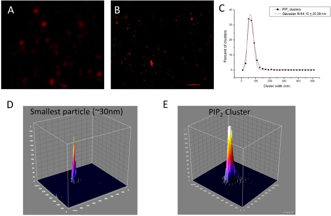
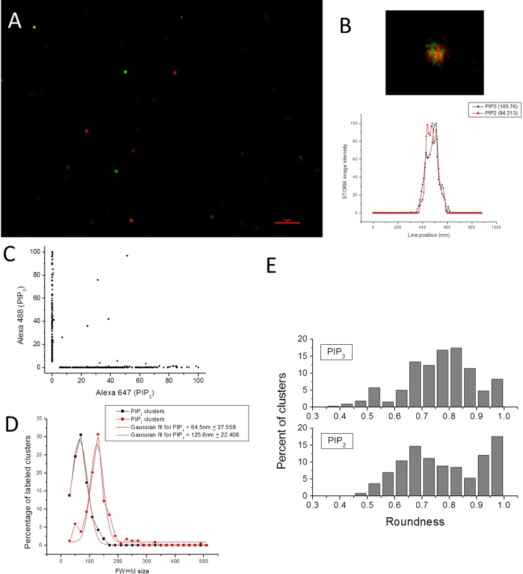
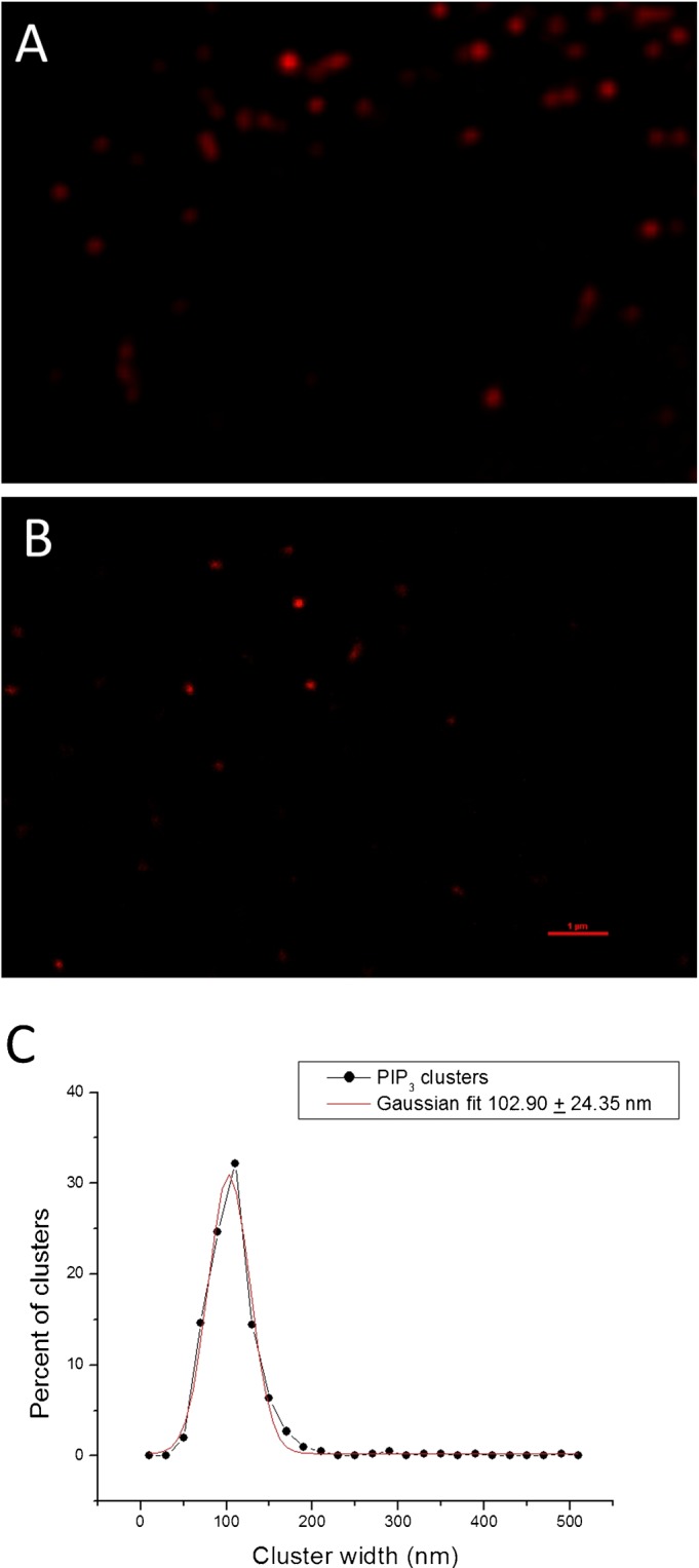
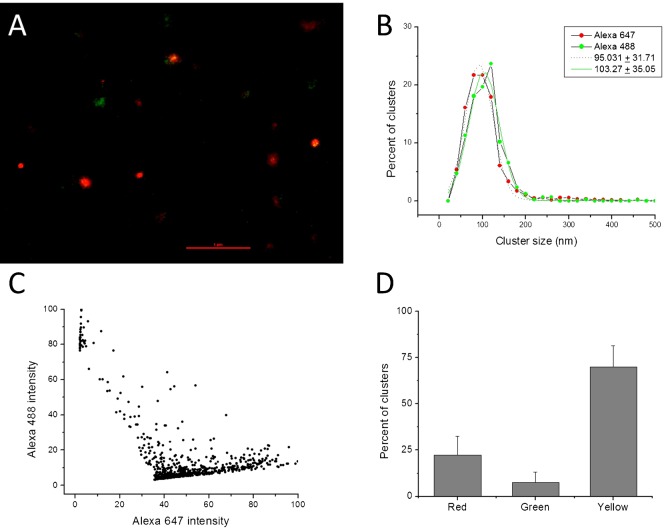
PIP(2) and PIP(3) are implicated in a wide variety of cellular signaling pathways at the plasma membrane. We have used STORM imaging to localize clusters of PIP(2) and PIP(3) to distinct nanoscale regions within the plasma membrane of PC12 cells. With anti-phospholipid antibodies directly conjugated with AlexaFluor 647, we found that PIP(2) clusters in membrane domains of 64.5±27.558 nm, while PIP(3) clusters had a size of 125.6±22.408 nm. With two color direct STORM imaging we show that >99% of phospholipid clusters have only one or other phospholipid present. These results indicate that lipid nano-domains can be readily identified using super-resolution imaging techniques, and that the lipid composition and size of clusters is tightly regulated.
PIP(2) 和 PIP(3) 参与了质膜上多种细胞信号通路。我们使用 STORM 成像技术将 PIP(2) 和 PIP(3) 簇定位于 PC12 细胞质膜内的不同纳米级区域。使用直接与 AlexaFluor 647 偶联的抗磷脂抗体,我们发现 PIP(2) 簇位于 64.5±27.558nm 的膜域中,而 PIP(3) 簇的大小为 125.6±22.408nm。通过双色直接 STORM 成像,我们表明 >99%的磷脂簇只存在一种或另一种磷脂。这些结果表明,使用超分辨率成像技术可以很容易地识别脂质纳米域,并且簇的脂质组成和大小受到严格调控。