Wang Nan, Shao Yeqing, Mei Yan, Zhang Li, Li Qinggang, Li Diangeng, Shi Suozhu, Hong Quan, Lin Hongli, Chen Xiangmei
Stem Cell Res Ther. 2012 Dec 6;3(6):51. doi: 10.1186/scrt142.
We previously found that mesenchymal stem cells (MSCs) injected intravenously could attenuate peritoneal adhesion by secreting tumor necrosis alpha-stimulating gene (TSG)-6, while MSCs injected intraperitoneally could not. However, the underlying mechanism remains unclear. This study was designed to investigate the means by which MSCs exert their effects.
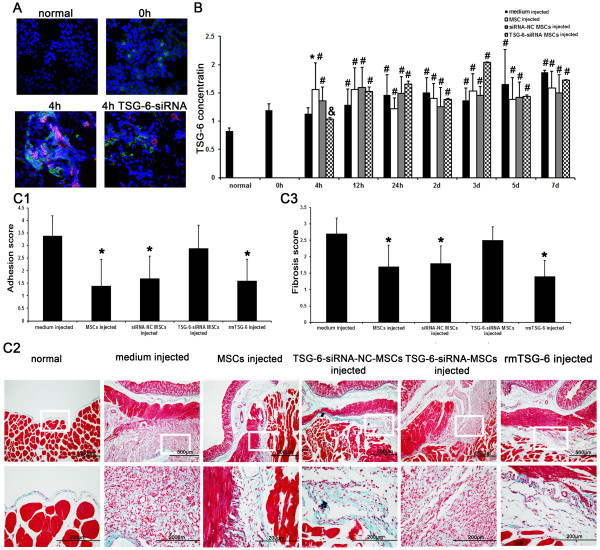
Rat bone marrow-derived MSCs/red fluorescent protein (RFP) were injected either intraperitoneally or intravenously into Sprague-Dawley (SD) rats at different time points after peritoneal scraping. Peritoneal adhesions were evaluated macroscopically at day 14 after scraping. The distribution of MSCs injected intraperitoneally or intravenously was traced by two-photon fluorescence confocal imaging and immunofluorescence microscopy. The co-localization of MSCs and macrophages in the lung and the spleen, and the expression of TSG-6 in MSCs trapped in the lung or the spleen were evaluated by immunofluorescence microscopy. The concentration of TSG-6 in serum was evaluated by ELISA. After intravenous injection of TSG-6- small interfering (si) RNA-MSCs, the expression of TSG-6 in MSCs and the concentration of TSG-6 in serum were reevaluated, and peritoneal adhesions were evaluated macroscopically and histologically.
MSCs injected intraperitoneally failed to reduce peritoneal adhesion, and MSCs injected intravenously markedly improved peritoneal adhesion. Two-photon fluorescence confocal imaging showed that MSCs injected intravenously accumulated mainly in the lung, where they remained for seven days, and immunofluorescence microscopy showed few MSCs phagocytosed by macrophages. In contrast, large numbers of MSCs accumulated in the spleen with obvious phagocytosis by macrophages even at 4 hours after intraperitoneal injection. Immunofluorescence microscopy showed that MSCs that accumulated in the lung after intravenous injection could express TSG-6 within 12 hours, but TSG-6-siRNA-MSCs or MSCs accumulated in the spleen after intraperitoneal injection did not. ELISA showed that the concentration of TSG-6 in serum was increased at 4 hours after intravenous injection of MSCs, while there was no increase after injection of TSG-6-siRNA-MSCs or after intraperitoneal injection of MSCs. Moreover, intravenous injection of TSG-6-siRNA-MSCs failed to attenuate peritoneal adhesion.
Our findings suggest that intravenously injected MSCs accumulated in the lung and attenuated peritoneal adhesion by secreting TSG-6, but intraperitoneally injected MSCs were phagocytosed by macrophages in the spleen and failed to attenuate peritoneal adhesion.
我们之前发现静脉注射间充质干细胞(MSCs)可通过分泌肿瘤坏死因子α刺激基因(TSG)-6减轻腹膜粘连,而腹腔注射MSCs则不能。然而,其潜在机制仍不清楚。本研究旨在探究MSCs发挥作用的方式。
在腹膜刮擦后的不同时间点,将大鼠骨髓来源的MSCs/红色荧光蛋白(RFP)腹腔内或静脉内注射到Sprague-Dawley(SD)大鼠体内。在刮擦后第14天宏观评估腹膜粘连情况。通过双光子荧光共聚焦成像和免疫荧光显微镜追踪腹腔内或静脉内注射的MSCs的分布。通过免疫荧光显微镜评估肺和脾中MSCs与巨噬细胞的共定位,以及被困在肺或脾中的MSCs中TSG-6的表达。通过酶联免疫吸附测定(ELISA)评估血清中TSG-6的浓度。静脉注射TSG-6小干扰(si)RNA-MSCs后,重新评估MSCs中TSG-6的表达和血清中TSG-6的浓度,并宏观和组织学评估腹膜粘连情况。
腹腔注射的MSCs未能减轻腹膜粘连,而静脉注射的MSCs显著改善了腹膜粘连。双光子荧光共聚焦成像显示,静脉注射的MSCs主要积聚在肺中,并在肺中停留7天,免疫荧光显微镜显示很少有MSCs被巨噬细胞吞噬。相反,即使在腹腔注射后4小时,大量MSCs积聚在脾中,巨噬细胞有明显的吞噬作用。免疫荧光显微镜显示,静脉注射后积聚在肺中的MSCs可在1十二小时内表达TSG-6,但TSG-6-siRNA-MSCs或腹腔注射后积聚在脾中的MSCs则不表达。ELISA显示,静脉注射MSCs后4小时血清中TSG-6的浓度升高,而注射TSG-6-siRNA-MSCs后或腹腔注射MSCs后则没有升高。此外,静脉注射TSG-6-siRNA-MSCs未能减轻腹膜粘连。
我们的研究结果表明,静脉注射的MSCs积聚在肺中并通过分泌TSG-6减轻腹膜粘连,但腹腔注射的MSCs被脾中的巨噬细胞吞噬且未能减轻腹膜粘连。