• 文献检索
  • 文档翻译
  • 深度研究
  • 学术资讯
  • Suppr Zotero 插件Zotero 插件
  • 邀请有礼
  • 套餐&价格
  • 历史记录
应用&插件
Suppr Zotero 插件Zotero 插件浏览器插件Mac 客户端Windows 客户端微信小程序
定价
高级版会员购买积分包购买API积分包
服务
文献检索文档翻译深度研究API 文档MCP 服务
关于我们
关于 Suppr公司介绍联系我们用户协议隐私条款
关注我们

Suppr 超能文献

核心技术专利:CN118964589B侵权必究
粤ICP备2023148730 号-1Suppr @ 2026

文献检索

告别复杂PubMed语法,用中文像聊天一样搜索,搜遍4000万医学文献。AI智能推荐,让科研检索更轻松。

立即免费搜索

文件翻译

保留排版,准确专业,支持PDF/Word/PPT等文件格式,支持 12+语言互译。

免费翻译文档

深度研究

AI帮你快速写综述,25分钟生成高质量综述,智能提取关键信息,辅助科研写作。

立即免费体验

有丝分裂细胞内的微管正端是具有锚定、信号和力偶联作用的“活动平台”。

Microtubule plus-ends within a mitotic cell are 'moving platforms' with anchoring, signalling and force-coupling roles.

机构信息

Department of Genetics, University of Cambridge, Downing Site, Downing Street, Cambridge CB2 3EH, UK.

出版信息

Open Biol. 2012 Nov;2(11):120132. doi: 10.1098/rsob.120132.

DOI:10.1098/rsob.120132
PMID:23226599
原文链接:https://pmc.ncbi.nlm.nih.gov/articles/PMC3513837/
Abstract

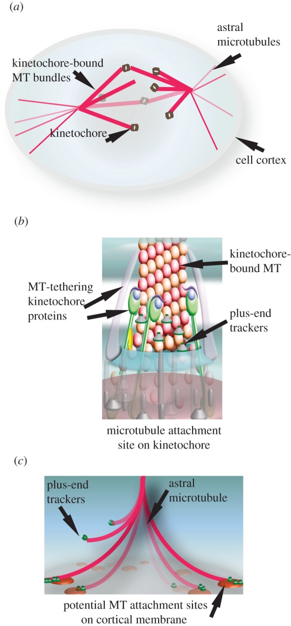
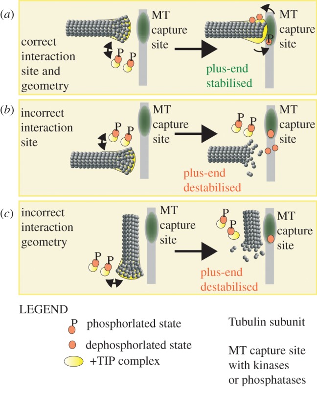
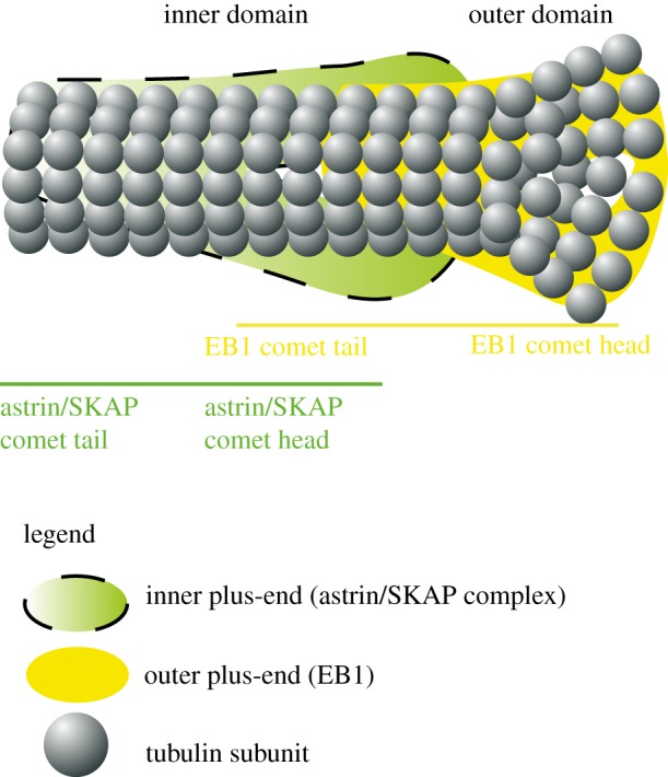
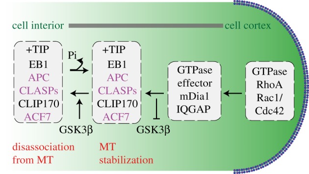
The microtubule polymer grows and shrinks predominantly from one of its ends called the 'plus-end'. Plus-end regulation during interphase is well understood. However, mitotic regulation of plus-ends is only beginning to be understood in mammalian cells. During mitosis, the plus-ends are tethered to specialized microtubule capture sites. At these sites, plus-end-binding proteins are loaded and unloaded in a regulated fashion. Proper tethering of plus-ends to specialized sites is important so that the microtubule is able to translate its growth and shrinkage into pushing and pulling forces that move bulky subcellular structures. We discuss recent advances on how mitotic plus-ends are tethered to distinct subcellular sites and how plus-end-bound proteins can modulate the forces that move subcellular structures. Using end binding 1 (EB1) as a prototype plus-end-binding protein, we highlight the complex network of plus-end-binding proteins and their regulation through phosphorylation. Finally, we develop a speculative 'moving platform' model that illustrates the plus-end's role in distinguishing correct versus incorrect microtubule interactions.

摘要

微管聚合物主要从其称为“正端”的一个末端生长和收缩。 间期的正端调控已经得到很好的理解。 然而,有丝分裂过程中对正端的调控才刚刚开始在哺乳动物细胞中被理解。 在有丝分裂过程中,正端被固定在专门的微管捕获位点上。 在这些位点上,正端结合蛋白以受调控的方式加载和卸载。 正端与专门的位点适当固定非常重要,这样微管才能将其生长和收缩转化为推动和拉动的力,从而移动体积较大的亚细胞结构。 我们讨论了有丝分裂正端与不同亚细胞位点固定的最新进展,以及正端结合蛋白如何调节移动亚细胞结构的力。 我们使用末端结合蛋白 1(EB1)作为正端结合蛋白的原型,强调了正端结合蛋白及其通过磷酸化调节的复杂网络。 最后,我们提出了一个推测性的“移动平台”模型,说明了正端在区分正确和错误的微管相互作用中的作用。

https://cdn.ncbi.nlm.nih.gov/pmc/blobs/a318/3513837/45992d74aee1/rsob-2-120132-g4.jpg
https://cdn.ncbi.nlm.nih.gov/pmc/blobs/a318/3513837/3764bb45cd74/rsob-2-120132-g1.jpg
https://cdn.ncbi.nlm.nih.gov/pmc/blobs/a318/3513837/df100894c095/rsob-2-120132-g2.jpg
https://cdn.ncbi.nlm.nih.gov/pmc/blobs/a318/3513837/e98aab383154/rsob-2-120132-g3.jpg
https://cdn.ncbi.nlm.nih.gov/pmc/blobs/a318/3513837/45992d74aee1/rsob-2-120132-g4.jpg

相似文献

1
Microtubule plus-ends within a mitotic cell are 'moving platforms' with anchoring, signalling and force-coupling roles.有丝分裂细胞内的微管正端是具有锚定、信号和力偶联作用的“活动平台”。
Open Biol. 2012 Nov;2(11):120132. doi: 10.1098/rsob.120132.
2
Distinct mechanisms govern the localisation of Drosophila CLIP-190 to unattached kinetochores and microtubule plus-ends.不同的机制控制果蝇CLIP-190定位于未附着的动粒和微管正端。
J Cell Sci. 2005 Aug 15;118(Pt 16):3781-90. doi: 10.1242/jcs.02504.
3
Regulation of a dynamic interaction between two microtubule-binding proteins, EB1 and TIP150, by the mitotic p300/CBP-associated factor (PCAF) orchestrates kinetochore microtubule plasticity and chromosome stability during mitosis.由有丝分裂 p300/CBP 相关因子(PCAF)调节的两个微管结合蛋白 EB1 和 TIP150 之间的动态相互作用,协调有丝分裂过程中着丝粒微管的可塑性和染色体的稳定性。
J Biol Chem. 2013 May 31;288(22):15771-85. doi: 10.1074/jbc.M112.448886. Epub 2013 Apr 17.
4
Optogenetic EB1 inactivation shortens metaphase spindles by disrupting cortical force-producing interactions with astral microtubules.光遗传学 EB1 失活通过破坏与星体微管的皮质力产生相互作用来缩短中期纺锤体。
Curr Biol. 2022 Mar 14;32(5):1197-1205.e4. doi: 10.1016/j.cub.2022.01.017. Epub 2022 Jan 31.
5
Mitotic regulator SKAP forms a link between kinetochore core complex KMN and dynamic spindle microtubules.有丝分裂调节因子 SKAP 连接着动粒核心复合物 KMN 和动态纺锤体微管。
J Biol Chem. 2012 Nov 16;287(47):39380-90. doi: 10.1074/jbc.M112.406652. Epub 2012 Oct 3.
6
EB1 targets to kinetochores with attached, polymerizing microtubules.EB1靶向附着有正在聚合的微管的动粒。
Mol Biol Cell. 2002 Dec;13(12):4308-16. doi: 10.1091/mbc.e02-04-0236.
7
EB1 acetylation by P300/CBP-associated factor (PCAF) ensures accurate kinetochore-microtubule interactions in mitosis.P300/CBP 相关因子(PCAF)对 EB1 的乙酰化作用确保了有丝分裂中着丝粒-微管相互作用的准确性。
Proc Natl Acad Sci U S A. 2012 Oct 9;109(41):16564-9. doi: 10.1073/pnas.1202639109. Epub 2012 Sep 21.
8
Kinetochore-microtubule attachment in human cells is regulated by the interaction of a conserved motif of Ska1 with EB1.人细胞中的着丝粒微管附着由 Ska1 的保守基序与 EB1 的相互作用调节。
J Biol Chem. 2023 Feb;299(2):102853. doi: 10.1016/j.jbc.2022.102853. Epub 2022 Dec 31.
9
The growth speed of microtubules with XMAP215-coated beads coupled to their ends is increased by tensile force.带有 XMAP215 涂层珠的微管末端的生长速度会因张力而增加。
Proc Natl Acad Sci U S A. 2013 Sep 3;110(36):14670-5. doi: 10.1073/pnas.1218053110. Epub 2013 Aug 20.
10
Regulation of mitotic spindle orientation by phosphorylation of end binding protein 1.通过磷酸化末端结合蛋白 1 调节有丝分裂纺锤体的取向。
Exp Cell Res. 2019 Nov 1;384(1):111618. doi: 10.1016/j.yexcr.2019.111618. Epub 2019 Sep 7.

引用本文的文献

1
Sliding of antiparallel microtubules drives bipolarization of monoastral spindles.对向微管的滑动驱动单星纺锤体的两极化。
Cytoskeleton (Hoboken). 2024 Feb-Mar;81(2-3):167-183. doi: 10.1002/cm.21800. Epub 2023 Oct 9.
2
Deep learning techniques and mathematical modeling allow 3D analysis of mitotic spindle dynamics.深度学习技术和数学建模允许对有丝分裂纺锤体动力学进行 3D 分析。
J Cell Biol. 2023 May 1;222(5). doi: 10.1083/jcb.202111094. Epub 2023 Mar 2.
3
The Small-Molecule Inhibitor MRIA9 Reveals Novel Insights into the Cell Cycle Roles of SIK2 in Ovarian Cancer Cells.

本文引用的文献

1
The kinetochore-bound Ska1 complex tracks depolymerizing microtubules and binds to curved protofilaments.着丝粒结合的 Ska1 复合物追踪去聚合的微管,并与弯曲的原纤维结合。
Dev Cell. 2012 Nov 13;23(5):968-80. doi: 10.1016/j.devcel.2012.09.012. Epub 2012 Oct 18.
2
Cdk1 and Plk1 mediate a CLASP2 phospho-switch that stabilizes kinetochore-microtubule attachments.Cdk1 和 Plk1 介导 CLASP2 的磷酸化开关,稳定着动粒-微管连接。
J Cell Biol. 2012 Oct 15;199(2):285-301. doi: 10.1083/jcb.201203091. Epub 2012 Oct 8.
3
The Ndc80 kinetochore complex directly modulates microtubule dynamics.
小分子抑制剂MRIA9揭示了SIK2在卵巢癌细胞细胞周期作用中的新见解。
Cancers (Basel). 2021 Jul 21;13(15):3658. doi: 10.3390/cancers13153658.
4
Epigenome-wide DNA methylation analysis of small cell lung cancer cell lines suggests potential chemotherapy targets.对小细胞肺癌细胞系的全基因组 DNA 甲基化分析表明了潜在的化疗靶点。
Clin Epigenetics. 2020 Jun 25;12(1):93. doi: 10.1186/s13148-020-00876-8.
5
Kinetochores attached to microtubule-ends are stabilised by Astrin bound PP1 to ensure proper chromosome segregation.着丝粒与微管末端相连,由 Astrin 结合 PP1 稳定,以确保染色体的正确分离。
Elife. 2019 Dec 6;8:e49325. doi: 10.7554/eLife.49325.
6
Mapping the kinetochore MAP functions required for stabilizing microtubule attachments to chromosomes during metaphase.绘制在有丝分裂中期稳定微管与染色体连接所必需的着丝粒 MAP 功能图。
Cytoskeleton (Hoboken). 2019 Jun;76(6):398-412. doi: 10.1002/cm.21559. Epub 2019 Sep 9.
7
Microtubules: A Key to Understand and Correct Neuronal Defects in CDKL5 Deficiency Disorder?微管:理解和纠正 CDKL5 缺乏症中神经元缺陷的关键?
Int J Mol Sci. 2019 Aug 21;20(17):4075. doi: 10.3390/ijms20174075.
8
Insulin Induces Microtubule Stabilization and Regulates the Microtubule Plus-end Tracking Protein Network in Adipocytes.胰岛素诱导脂肪细胞微管稳定并调节微管末端追踪蛋白网络。
Mol Cell Proteomics. 2019 Jul;18(7):1363-1381. doi: 10.1074/mcp.RA119.001450. Epub 2019 Apr 24.
9
Spindle rotation in human cells is reliant on a MARK2-mediated equatorial spindle-centering mechanism.纺锤体在人类细胞中的旋转依赖于 MARK2 介导的赤道纺锤体定中机制。
J Cell Biol. 2018 Sep 3;217(9):3057-3070. doi: 10.1083/jcb.201804166. Epub 2018 Jun 25.
10
Sodium Channel Remodeling in Subcellular Microdomains of Murine Failing Cardiomyocytes.鼠衰竭心肌细胞亚细胞微域中的钠通道重构。
J Am Heart Assoc. 2017 Dec 8;6(12):e007622. doi: 10.1161/JAHA.117.007622.
Ndc80 动粒复合物直接调节微管动力学。
Proc Natl Acad Sci U S A. 2012 Oct 2;109(40):16113-8. doi: 10.1073/pnas.1209615109. Epub 2012 Aug 20.
4
A Proteome-wide screen for mammalian SxIP motif-containing microtubule plus-end tracking proteins.哺乳动物 SxIP 基序包含的微管正极追踪蛋白的蛋白质组范围筛选。
Curr Biol. 2012 Oct 9;22(19):1800-7. doi: 10.1016/j.cub.2012.07.047. Epub 2012 Aug 9.
5
Phosphoregulation of STIM1 leads to exclusion of the endoplasmic reticulum from the mitotic spindle.STIM1 的磷酸化调节导致内质网从有丝分裂纺锤体中排除。
Curr Biol. 2012 Aug 21;22(16):1487-93. doi: 10.1016/j.cub.2012.05.057. Epub 2012 Jun 28.
6
Mechanism for astral microtubule capture by cortical Bud6p priming spindle polarity in S. cerevisiae.星状微管通过 Bud6p 对皮层的预定位捕获机制来决定酵母的纺锤体极性。
Curr Biol. 2012 Jun 19;22(12):1075-83. doi: 10.1016/j.cub.2012.04.059. Epub 2012 May 17.
7
Kif18A and chromokinesins confine centromere movements via microtubule growth suppression and spatial control of kinetochore tension.Kif18A 和染色质运动蛋白通过抑制微管生长和动粒张力的空间控制来限制着丝粒运动。
Dev Cell. 2012 May 15;22(5):1017-29. doi: 10.1016/j.devcel.2012.02.013.
8
EBs recognize a nucleotide-dependent structural cap at growing microtubule ends.EBs 识别生长中的微管末端依赖核苷酸的结构帽。
Cell. 2012 Apr 13;149(2):371-82. doi: 10.1016/j.cell.2012.02.049.
9
Multisite phosphorylation disrupts arginine-glutamate salt bridge networks required for binding of cytoplasmic linker-associated protein 2 (CLASP2) to end-binding protein 1 (EB1).多位点磷酸化破坏了精氨酸-谷氨酸盐桥网络,这些网络对于细胞质连接蛋白 2 (CLASP2)与末端结合蛋白 1 (EB1)的结合是必需的。
J Biol Chem. 2012 May 18;287(21):17050-17064. doi: 10.1074/jbc.M111.316661. Epub 2012 Mar 29.
10
Dynein tethers and stabilizes dynamic microtubule plus ends.动力蛋白将和稳定动态微管的正端。
Curr Biol. 2012 Apr 10;22(7):632-7. doi: 10.1016/j.cub.2012.02.023. Epub 2012 Mar 22.