Department of Molecular Cell Biology, Leiden University Medical Center, PO Box 9600, 2300RC Leiden, The Netherlands.
J Synchrotron Radiat. 2013 Jan;20(Pt 1):58-66. doi: 10.1107/S0909049512044408. Epub 2012 Nov 29.
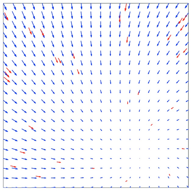
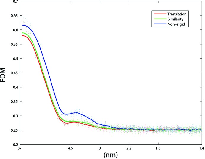
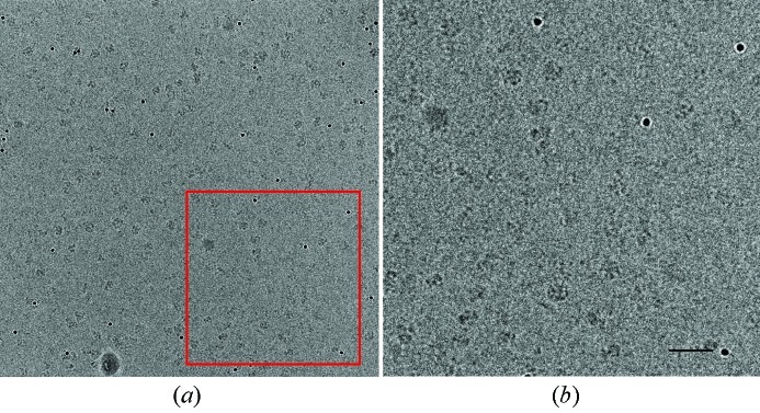
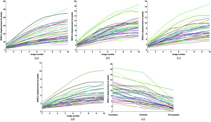
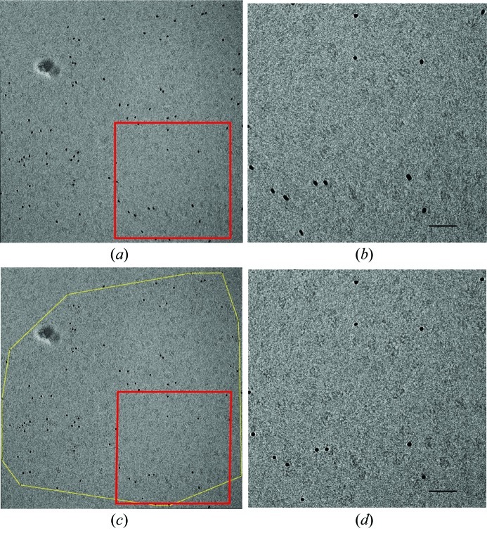
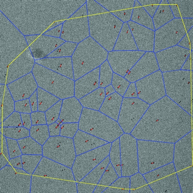
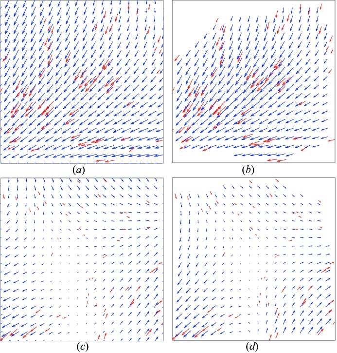
The typical dose used to record cryo-electron microscopy images from vitrified biological specimens is so high that radiation-induced structural alterations are bound to occur during data acquisition. Integration of all scattered electrons into one image can lead to significant blurring, particularly if the data are collected from an unsupported thin layer of ice suspended over the holes of a support film. Here, the dose has been fractioned and exposure series have been acquired in order to study beam-induced specimen movements under low dose conditions, prior to bubbling. Gold particles were added to the protein sample as fiducial markers. These were automatically localized and tracked throughout the exposure series and showed correlated motions within small patches, with larger amplitudes of motion vectors at the start of a series compared with the end of each series. A non-rigid scheme was used to register all images within each exposure series, using natural neighbor interpolation with the gold particles as anchor points. The procedure increases the contrast and resolution of the examined macromolecules.
用于记录玻璃化生物样本的冷冻电子显微镜图像的典型剂量如此之高,以至于在数据采集过程中必然会发生辐射诱导的结构改变。将所有散射电子整合到一个图像中会导致明显的模糊,特别是如果数据是从悬浮在支撑膜孔上的无支撑薄冰层中收集的。在这里,为了在冒泡之前研究低剂量条件下的束引起的样品运动,已经对剂量进行了分段,并获取了一系列暴露。金颗粒被添加到蛋白质样品中作为基准标记。这些标记物在整个暴露系列中自动定位和跟踪,并在小区域内显示出相关的运动,与每个系列的末尾相比,在系列的开始时运动矢量的幅度更大。使用自然邻点插值和金颗粒作为锚点,使用非刚性方案对每个暴露系列中的所有图像进行配准。该过程增加了被检查的大分子的对比度和分辨率。