Institut de Recherche Expérimentale et Clinique, Pôle d'Imagerie Moléculaire, Radiothérapie et Oncologie and Pôle de Recherche Cardiovasculaire, Université Catholique de Louvain, Brussels, 1200, Belgium.
EJNMMI Res. 2012 Dec 28;2(1):64. doi: 10.1186/2191-219X-2-64.
There is a growing interest in developing non-invasive imaging techniques permitting infarct size (IS) measurements in mice. The aim of this study was to validate the high-resolution rodent Linoview single photon emission computed tomography (SPECT) system for non-invasive measurements of IS in mice by using a novel algorithm independent of a normal database, in comparison with histology.
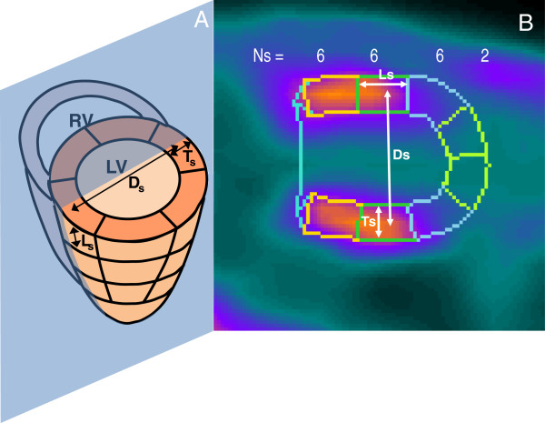
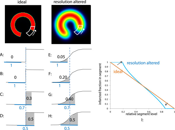
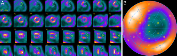
Eleven mice underwent a left coronary artery ligature. Seven days later, animals were imaged on the SPECT 2h30 after injection of 173 ± 27 MBq of Tc-99m-sestamibi. Mice were subsequently killed, and their hearts were excised for IS determination with triphenyltetrazolium chloride (TTC) staining. SPECT images were reconstructed using the expectation maximization maximum likelihood algorithm, and the IS was calculated using a novel algorithm applied on the 20-segment polar map provided by the commercially available QPS software (Cedars-Sinai Medical Center, CA, USA). This original method is attractive by the fact that it does not require the implementation of a normal perfusion database.
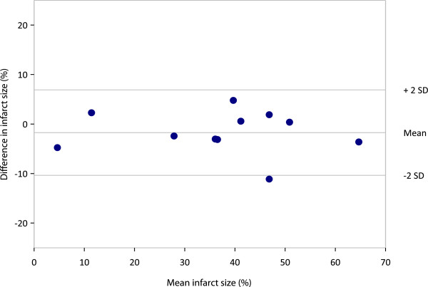
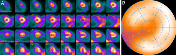
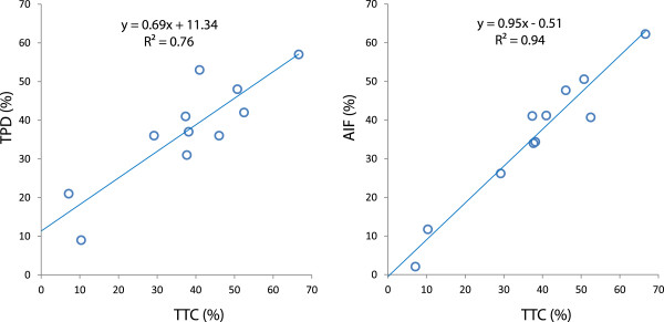
Reconstructed images allowed a clear delineation of the left ventricles borders in all mice. No significant difference was found between mean IS determined by SPECT and by TTC staining [37.9 ± 17.5% vs 35.6 ± 17.2%, respectively (P = 0.10)]. Linear regression analysis showed an excellent correlation between IS measured on the SPECT images and IS obtained with TTC staining (y = 0.95x + 0.03 (r = 0.97; P < 0.0001)), without bias, as demonstrated by the Bland-Altman plot.
Our results demonstrate the accuracy of the method for the measurement of myocardial IS in mice with the Linoview SPECT system.
人们对于开发能够无创测量小鼠梗死面积(IS)的影像学技术的兴趣日益浓厚。本研究旨在通过使用一种新的、不依赖正常数据库的算法,与组织学方法进行比较,验证新型高分辨率 Linoview 单光子发射计算机断层扫描(SPECT)系统用于非侵入性测量小鼠 IS 的准确性。
11 只小鼠进行左冠状动脉结扎。7 天后,在注射 173 ± 27 MBq 的 Tc-99m- sestamibi 后 2h30 对动物进行 SPECT 成像。随后处死小鼠,取出心脏,用氯化三苯基四氮唑(TTC)染色法测定 IS。使用期望最大化最大似然算法重建 SPECT 图像,并使用商业化 QPS 软件(美国加利福尼亚州雪松西奈医疗中心)提供的 20 节极坐标图上的新算法计算 IS。该原始方法的优点是它不需要实施正常灌注数据库。
重建后的图像能够清晰地描绘出所有小鼠的左心室边界。SPECT 测定的平均 IS 与 TTC 染色法测定的 IS 之间无显著差异[分别为 37.9 ± 17.5%和 35.6 ± 17.2%(P = 0.10)]。线性回归分析显示,SPECT 图像上测量的 IS 与 TTC 染色法获得的 IS 之间具有极好的相关性(y = 0.95x + 0.03(r = 0.97;P < 0.0001)),Bland-Altman 图表明没有偏差。
我们的结果表明,Linoview SPECT 系统在测量小鼠心肌 IS 方面具有很高的准确性。