Institute of Clinical Medicine and Department of Cardiology, Renmin Hospital, Hubei University of Medicine, Shiyan, Hubei, People's Republic of China.
PLoS One. 2012;7(12):e52537. doi: 10.1371/journal.pone.0052537. Epub 2012 Dec 28.
Poor survival of mesenchymal stem cells (MSC) compromised the efficacy of stem cell therapy for ischemic diseases. The aim of this study is to investigate the role of PEP-1-CAT transduction in MSC survival and its effect on ischemia-induced angiogenesis.
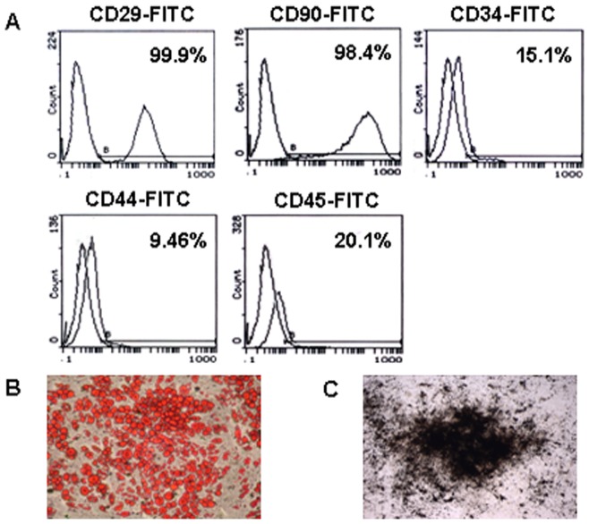
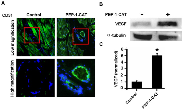
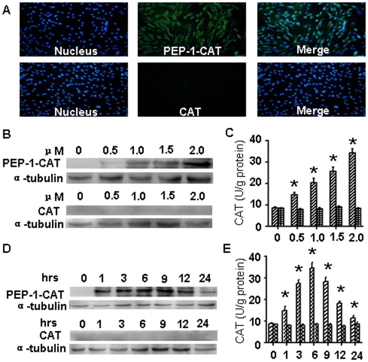
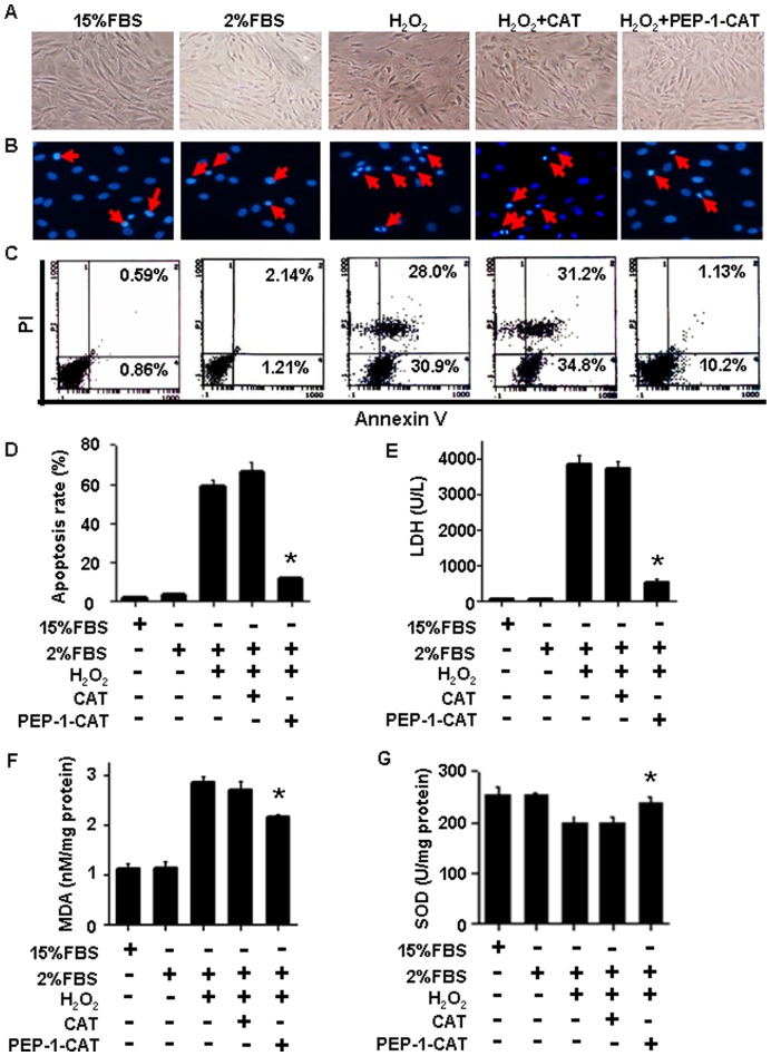
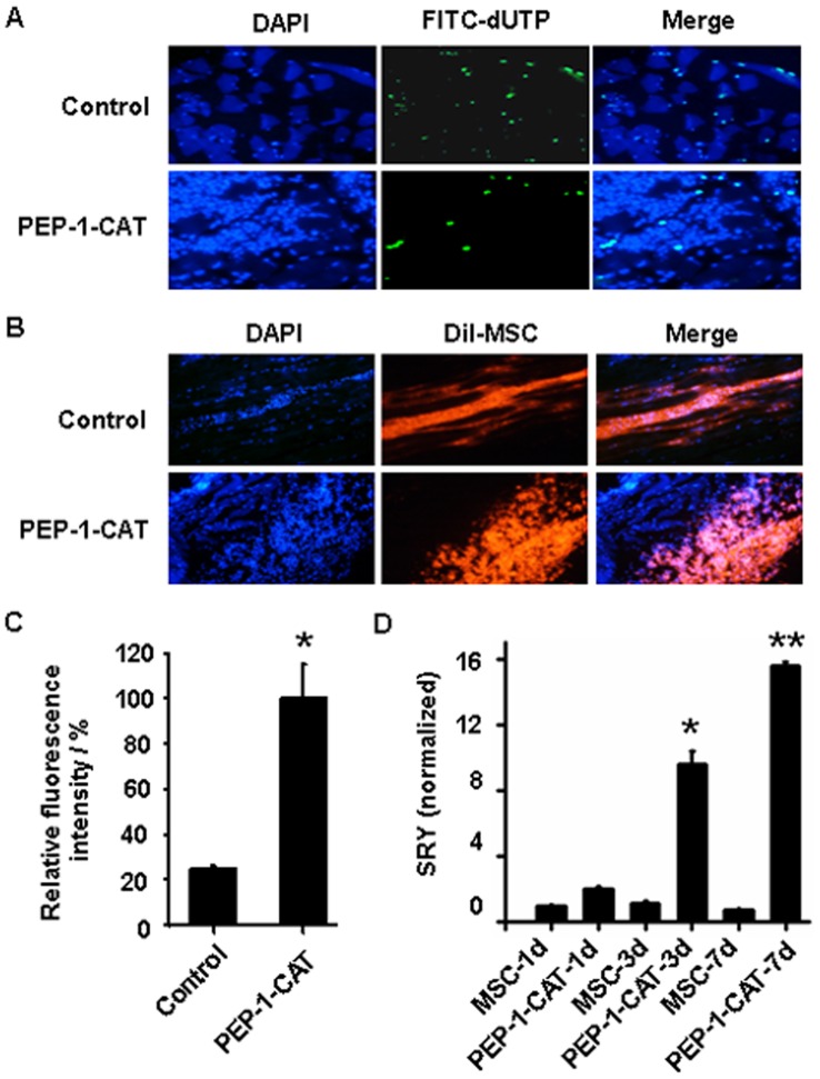
MSC apoptosis was evaluated by DAPI staining and quantified by Annexin V and PI double staining and Flow Cytometry. Malondialdehyde (MDA) content, lactate dehydrogenase (LDH) release, and Superoxide Dismutase (SOD) activities were simultaneously measured. MSC mitochondrial membrane potential was analyzed with JC-1 staining. MSC survival in rat muscles with gender-mismatched transplantation of the MSC after lower limb ischemia was assessed by detecting SRY expression. MSC apoptosis in ischemic area was determined by TUNEL assay. The effect of PEP-1-CAT-transduced MSC on angiogenesis in vivo was determined in the lower limb ischemia model.
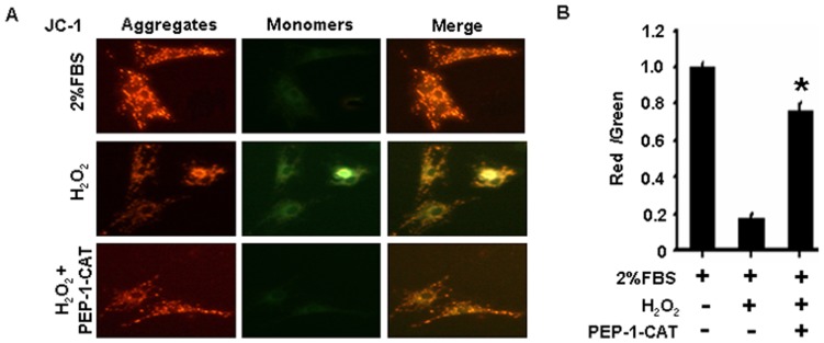
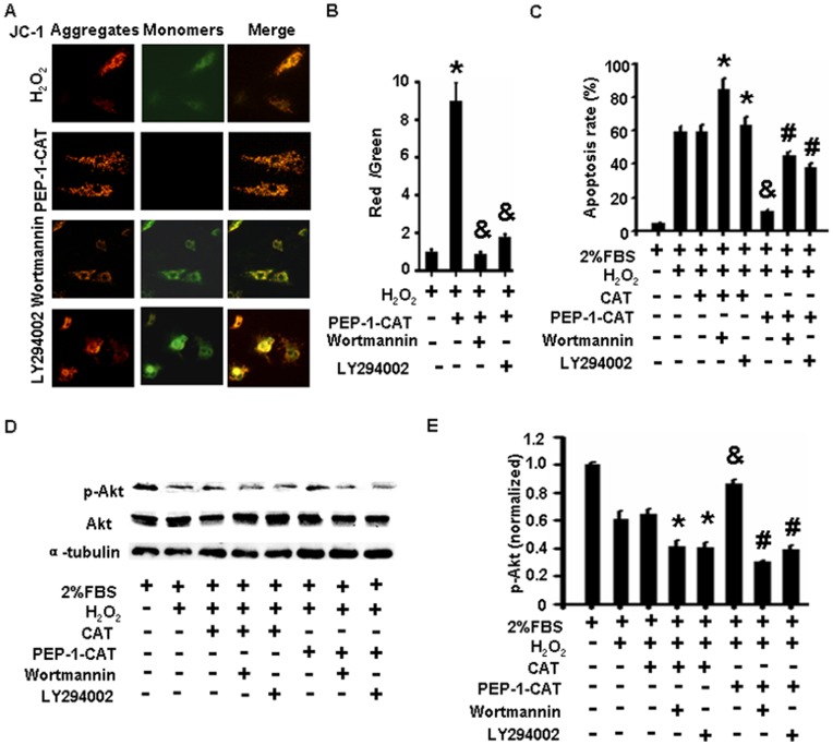
PEP-1-CAT transduction decreased MSC apoptosis rate while down-regulating MDA content and blocking LDH release as compared to the treatment with H(2)O(2) or CAT. However, SOD activity was up-regulated in PEP-1-CAT-transduced cells. Consistent with its effect on MSC apoptosis, PEP-1-CAT restored H(2)O(2)-attenuated mitochondrial membrane potential. Mechanistically, PEP-1-CAT blocked H(2)O(2)-induced down-regulation of PI3K/Akt activity, an essential signaling pathway regulating MSC apoptosis. In vivo, the viability of MSC implanted into ischemic area in lower limb ischemia rat model was increased by four-fold when transduced with PEP-1-CAT. Importantly, PEP-1-CAT-transduced MSC significantly enhanced ischemia-induced angiogenesis by up-regulating VEGF expression.
PEP-1-CAT-transduction was able to increase MSC viability by regulating PI3K/Akt activity, which stimulated ischemia-induced angiogenesis.
间充质干细胞(MSC)的存活率低,降低了干细胞疗法治疗缺血性疾病的疗效。本研究旨在探讨 PEP-1-CAT 转导在 MSC 存活中的作用及其对缺血诱导血管生成的影响。
通过 DAPI 染色评估 MSC 凋亡,并通过 Annexin V 和 PI 双重染色和流式细胞术定量。同时测量丙二醛(MDA)含量、乳酸脱氢酶(LDH)释放和超氧化物歧化酶(SOD)活性。用 JC-1 染色分析 MSC 线粒体膜电位。通过检测 SRY 表达评估大鼠下肢缺血后 MSC 性别错配移植中 MSC 在肌肉中的存活情况。通过 TUNEL 检测法确定缺血区 MSC 的凋亡情况。在下肢缺血模型中,通过检测血管生成来确定 PEP-1-CAT 转导 MSC 的作用。
与 H 2 O 2 或 CAT 处理相比,PEP-1-CAT 转导降低了 MSC 凋亡率,同时下调 MDA 含量并阻断 LDH 释放。然而,PEP-1-CAT 转导的细胞中 SOD 活性上调。与对 MSC 凋亡的影响一致,PEP-1-CAT 恢复了 H 2 O 2 减弱的线粒体膜电位。在机制上,PEP-1-CAT 阻断了 H 2 O 2 诱导的 PI3K/Akt 活性下调,该活性是调节 MSC 凋亡的必需信号通路。在体内,当用 PEP-1-CAT 转导时,植入下肢缺血大鼠模型缺血区的 MSC 的存活率增加了四倍。重要的是,PEP-1-CAT 转导的 MSC 通过上调 VEGF 表达显著增强了缺血诱导的血管生成。
PEP-1-CAT 转导通过调节 PI3K/Akt 活性增加 MSC 活力,从而刺激缺血诱导的血管生成。