Institute of Clinical Medicine, Renmin Hospital, Hubei University of Medicine, Shiyan, Hubei 442000, China.
J Transl Med. 2013 May 6;11:113. doi: 10.1186/1479-5876-11-113.
Catalase (CAT) breaks down H2O2 into H2O and O2 to protects cells from oxidative damage. However, its translational potential is limited because exogenous CAT cannot enter living cells automatically. This study is aimed to investigate if PEP-1-CAT fusion protein can effectively protect cardiomyocytes from oxidative stress due to hypoxia/reoxygenation (H/R)-induced injury.
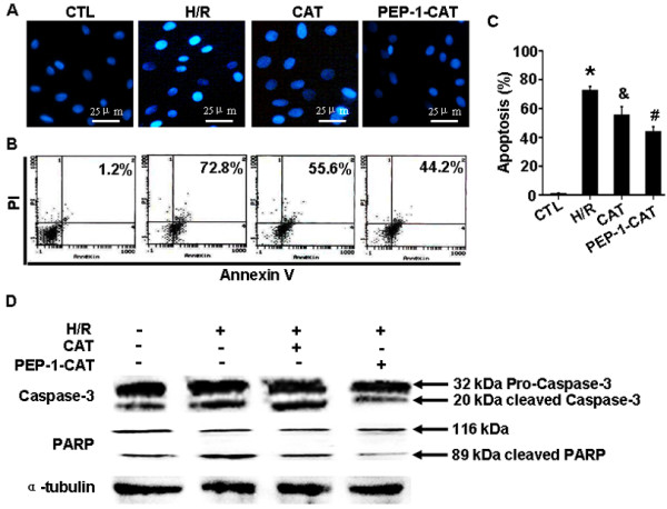
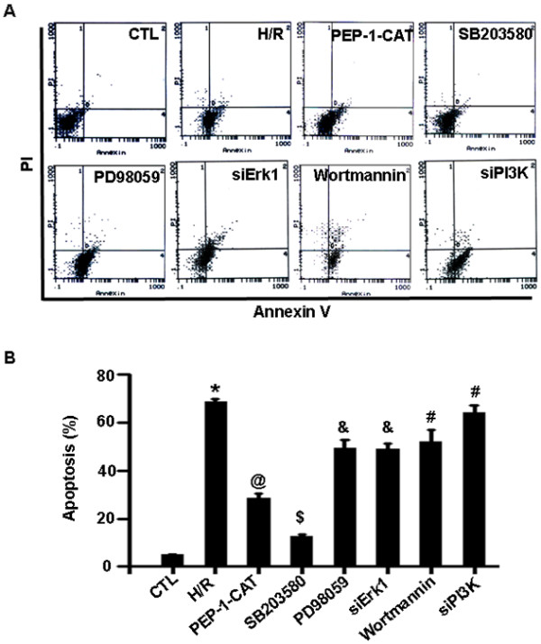
H9c2 cardomyocytes were pretreated with catalase (CAT) or PEP-1-CAT fusion protein followed by culturing in a hypoxia and re-oxygenation condition. Cell apoptosis were measured by Annexin V and PI double staining and Flow cytometry. Intracellular superoxide anion level was determined, and mitochondrial membrane potential was measured. Expression of apoptosis-related proteins including Bcl-2, Bax, Caspase-3, PARP, p38 and phospho-p38 was analyzed by western blotting.
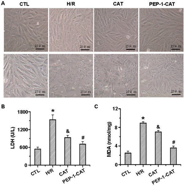
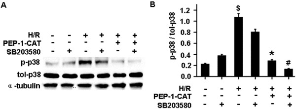
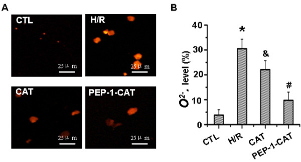
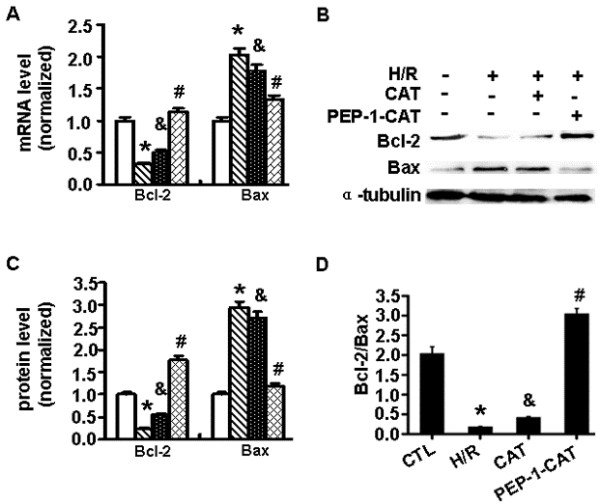
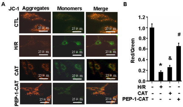
PEP-1-CAT protected H9c2 from H/R-induced morphological alteration and reduced the release of lactate dehydrogenase (LDH) and malondialdehyde content. Superoxide anion production was also decreased. In addition, PEP-1-CAT inhibited H9c2 apoptosis and blocked the expression of apoptosis stimulator Bax while increased the expression of Bcl-2, leading to an increased mitochondrial membrane potential. Mechanistically, PEP-1-CAT inhibited p38 MAPK while activating PI3K/Akt and Erk1/2 signaling pathways, resulting in blockade of Bcl2/Bax/mitochondrial apoptotic pathway.
Our study has revealed a novel mechanism by which PEP-1-CAT protects cardiomyocyte from H/R-induced injury. PEP-1-CAT blocks Bcl2/Bax/mitochondrial apoptotic pathway by inhibiting p38 MAPK while activating PI3K/Akt and Erk1/2 signaling pathways.
过氧化氢酶 (CAT) 将 H2O2 分解为 H2O 和 O2,以保护细胞免受氧化损伤。然而,由于外源性 CAT 不能自动进入活细胞,其翻译潜力受到限制。本研究旨在探讨 PEP-1-CAT 融合蛋白是否能有效保护心肌细胞免受缺氧/复氧 (H/R) 诱导损伤引起的氧化应激。
用 CAT 或 PEP-1-CAT 融合蛋白预处理 H9c2 心肌细胞,然后在缺氧和复氧条件下培养。用 Annexin V 和 PI 双重染色和流式细胞术检测细胞凋亡。测定细胞内超氧阴离子水平,测量线粒体膜电位。用 Western blot 分析凋亡相关蛋白包括 Bcl-2、Bax、Caspase-3、PARP、p38 和磷酸化 p38 的表达。
PEP-1-CAT 可保护 H9c2 免受 H/R 诱导的形态改变,并减少乳酸脱氢酶 (LDH) 的释放和丙二醛含量。超氧阴离子的产生也减少了。此外,PEP-1-CAT 抑制 H9c2 凋亡,阻止凋亡刺激剂 Bax 的表达,同时增加 Bcl-2 的表达,导致线粒体膜电位增加。机制上,PEP-1-CAT 抑制 p38 MAPK,同时激活 PI3K/Akt 和 Erk1/2 信号通路,从而阻断 Bcl2/Bax/线粒体凋亡途径。
本研究揭示了 PEP-1-CAT 保护心肌细胞免受 H/R 诱导损伤的新机制。PEP-1-CAT 通过抑制 p38 MAPK 而激活 PI3K/Akt 和 Erk1/2 信号通路,阻断 Bcl2/Bax/线粒体凋亡途径。