Bhattacharya Arnab, Dhar Pushpa, Mehra Raj D
Department of Anatomy, All India Institute of Medical Sciences, New Delhi, India.
Anat Cell Biol. 2012 Dec;45(4):229-40. doi: 10.5115/acb.2012.45.4.229. Epub 2012 Dec 14.
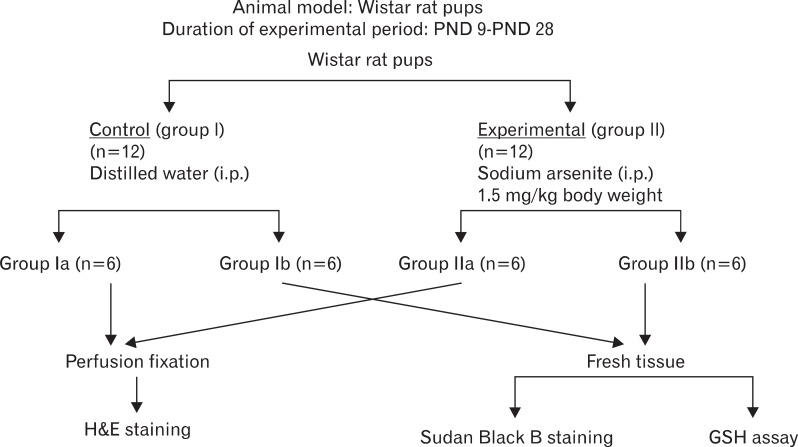
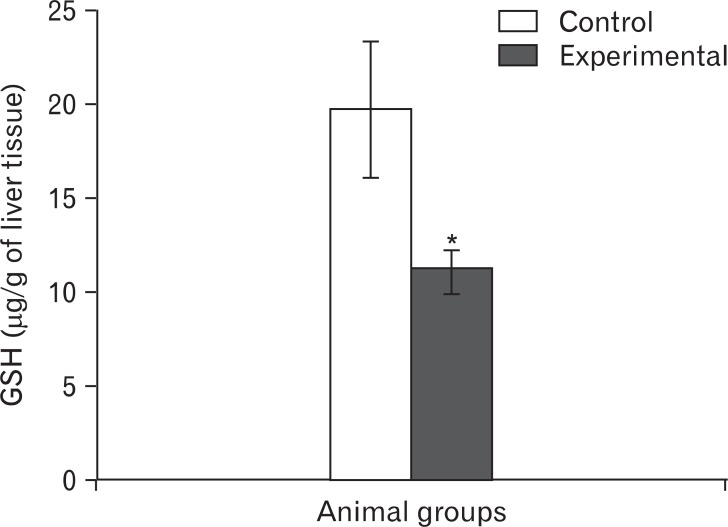
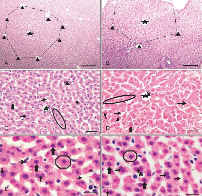
The effects of sodium arsenite exposure on the hepatic maturation period of cellular and functional reorganization in developing rat livers were evaluated. Animals received intraperitoneal injections of sodium arsenite (1.5 mg/kg body weight) or distilled water on days 9 to 28 after birth. On day 29, the animals were sacrificed either by cervical dislocation or by perfusion fixation. The perfusion fixed liver tissue was processed for paraffin embedding, sectioning and hematoxylin and eosin staining. The fresh liver tissue was processed for cryo-sectioning followed by Sudan Black B staining and for biochemical estimation of reduced glutathione. Microscopic observation revealed comparable preserved hepatic lobular patterns and distributions of uninucleate and binucleate hepatocytes in the control and the experimental groups. The mean nuclear area and diameter of the hepatocytes was increased in the experimental group. Lipid droplet distribution pattern in Sudan Black B stained sections revealed higher staining intensity towards the centrilobular area in both groups. Semiquantitative estimation of staining intensity showed lower mean gray values in zone 3 than in zones 2 and 1 (suggestive of the setting in of the adult pattern) in both groups. The reduced glutathione levels in the liver tissue and the altered nuclear size of the hepatocytes in the experimental group suggested the impairment of morphological and biochemical processes induced by arsenic exposure during the postnatal period.
评估了亚砷酸钠暴露对发育中大鼠肝脏细胞和功能重组肝成熟期的影响。在出生后第9至28天,动物接受腹腔注射亚砷酸钠(1.5 mg/kg体重)或蒸馏水。在第29天,通过颈椎脱臼或灌注固定处死动物。将灌注固定的肝组织进行石蜡包埋、切片及苏木精和伊红染色。将新鲜肝组织进行冷冻切片,随后进行苏丹黑B染色,并对还原型谷胱甘肽进行生化测定。显微镜观察显示,对照组和实验组的肝小叶模式以及单核和双核肝细胞的分布保存情况相当。实验组肝细胞的平均核面积和直径增加。苏丹黑B染色切片中的脂滴分布模式显示,两组中央小叶区域的染色强度均较高。染色强度的半定量估计显示,两组的3区平均灰度值均低于2区和1区(提示成人模式的形成)。实验组肝组织中还原型谷胱甘肽水平及肝细胞核大小的改变表明,出生后砷暴露会损害形态和生化过程。