Kajić Vedran, Esmaeelpour Marieh, Glittenberg Carl, Kraus Martin F, Honegger Joachim, Othara Richu, Binder Susanne, Fujimoto James G, Drexler Wolfgang
Center for Medical Physics and Biomedical Engineering, Medical University Vienna, General Hospital Vienna 4L, Waehringer Guertel 18-20, A-1090 Vienna, Austria.
Biomed Opt Express. 2013 Jan 1;4(1):134-50. doi: 10.1364/BOE.4.000134. Epub 2012 Dec 17.
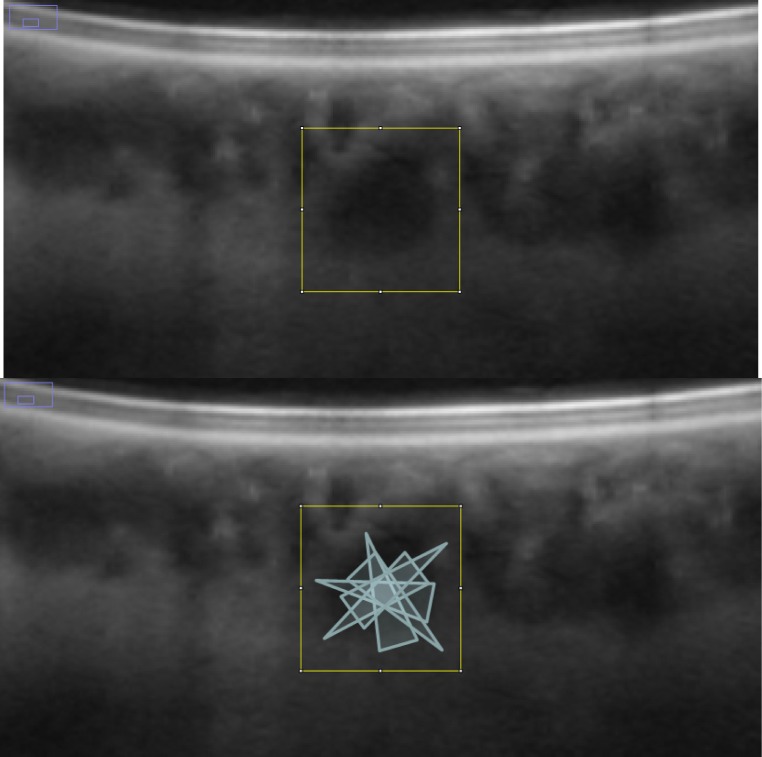
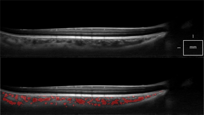
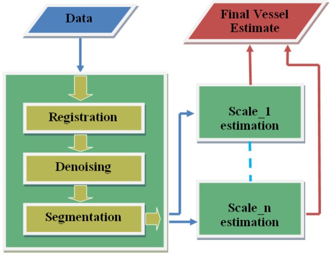
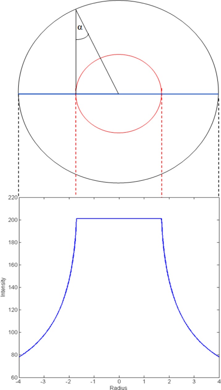
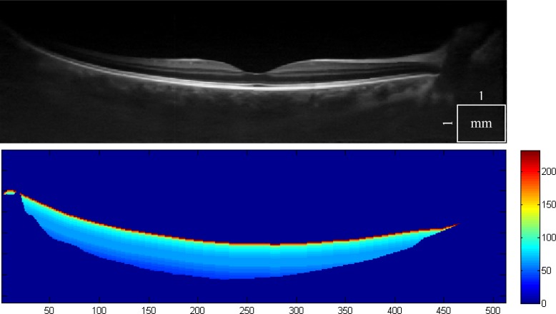
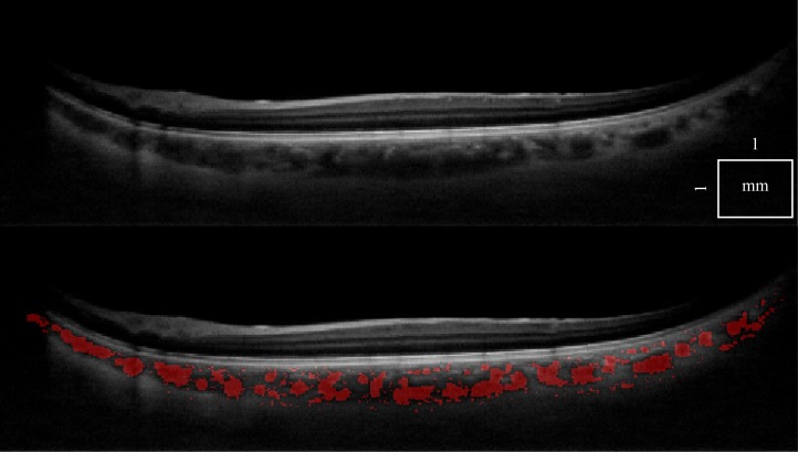
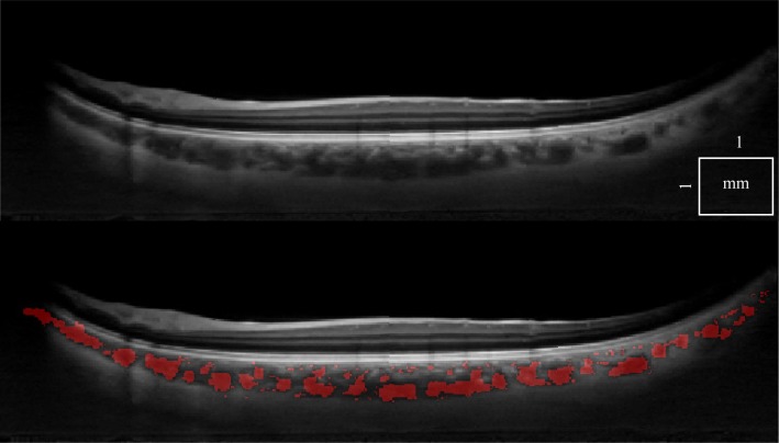
A fully automated, robust vessel segmentation algorithm has been developed for choroidal OCT, employing multiscale 3D edge filtering and projection of "probability cones" to determine the vessel "core", even in the tomograms with low signal-to-noise ratio (SNR). Based on the ideal vessel response after registration and multiscale filtering, with computed depth related SNR, the vessel core estimate is dilated to quantify the full vessel diameter. As a consequence, various statistics can be computed using the 3D choroidal vessel information, such as ratios of inner (smaller) to outer (larger) choroidal vessels or the absolute/relative volume of choroid vessels. Choroidal vessel quantification can be displayed in various forms, focused and averaged within a special region of interest, or analyzed as the function of image depth. In this way, the proposed algorithm enables unique visualization of choroidal watershed zones, as well as the vessel size reduction when investigating the choroid from the sclera towards the retinal pigment epithelium (RPE). To the best of our knowledge, this is the first time that an automatic choroidal vessel segmentation algorithm is successfully applied to 1060 nm 3D OCT of healthy and diseased eyes.
已开发出一种用于脉络膜光学相干断层扫描(OCT)的全自动、强大的血管分割算法,该算法采用多尺度3D边缘滤波和“概率锥”投影来确定血管“核心”,即使在信噪比(SNR)较低的断层图像中也能做到。基于配准和多尺度滤波后的理想血管响应,并结合计算出的与深度相关的SNR,对血管核心估计值进行扩张以量化整个血管直径。因此,可以使用3D脉络膜血管信息计算各种统计数据,例如脉络膜内(较小)血管与外(较大)血管的比例或脉络膜血管的绝对/相对体积。脉络膜血管量化可以以各种形式显示,在特殊感兴趣区域内聚焦并求平均值,或者作为图像深度的函数进行分析。通过这种方式,所提出的算法能够独特地可视化脉络膜分水岭区域,以及在从巩膜向视网膜色素上皮(RPE)研究脉络膜时血管尺寸的减小。据我们所知,这是首次将自动脉络膜血管分割算法成功应用于健康和患病眼睛的1060 nm 3D OCT。