Macromolecular X-Ray Crystallography Group, Structural Biology and Biophysics, Institute of Biotechnology, University of Helsinki Helsinki, Finland ; The National Doctoral Program in Informational and Structural Biology, Åbo Academy Turku, Finland.
Front Cell Infect Microbiol. 2013 Jan 8;2:169. doi: 10.3389/fcimb.2012.00169. eCollection 2012.
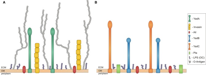
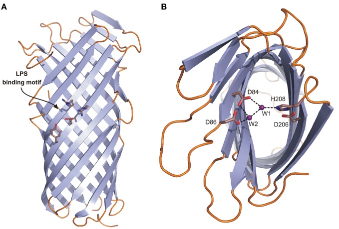
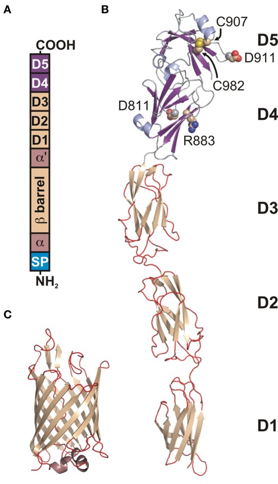
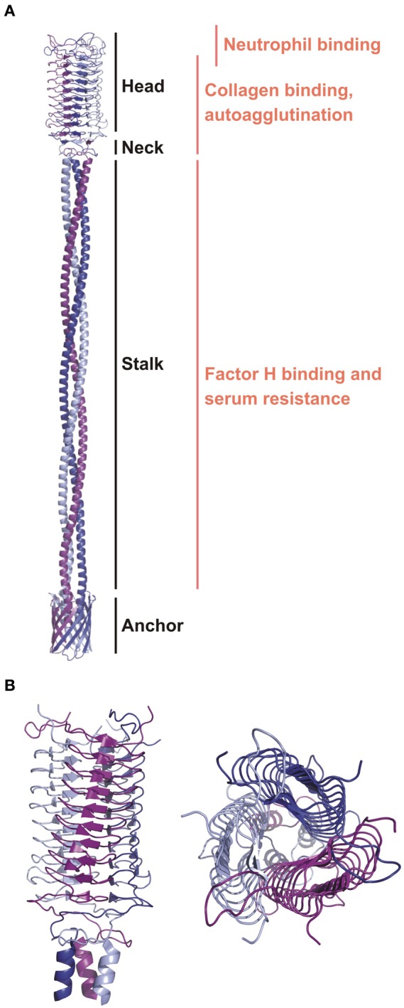
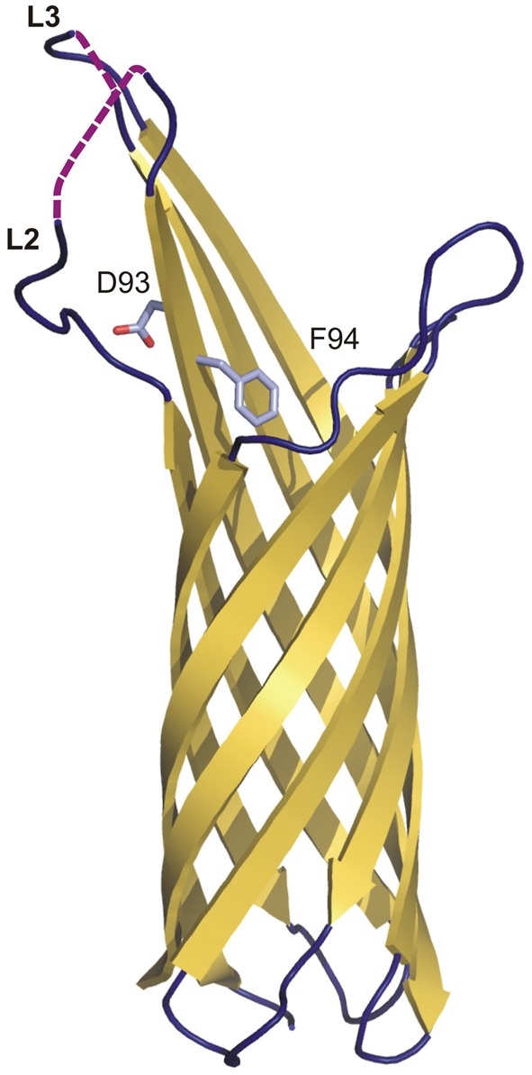
Among the seventeen species of the Gram-negative genus Yersinia, three have been shown to be virulent and pathogenic to humans and animals-Y. enterocolitica, Y. pseudotuberculosis, and Y. pestis. In order to be so, they are armoured with various factors that help them adhere to tissues and organelles, cross the cellular barrier and escape the immune system during host invasion. The group of proteins that mediate pathogen-host interactions constitute adhesins. Invasin, Ail, YadA, YadB, YadC, Pla, and pH 6 antigen belong to the most prominent and best-known Yersinia adhesins. They act at different times and stages of infection complementing each other by their ability to bind a variety of host molecules such as collagen, fibronectin, laminin, β1 integrins, and complement regulators. All the proteins are anchored in the bacterial outer membrane (OM), often forming rod-like or fimbrial-like structures that protrude to the extracellular milieu. Structural studies have shown that the anchor region forms a β-barrel composed of 8, 10, or 12 antiparallel β-strands. Depending on the protein, the extracellular part can be composed of several domains belonging to the immunoglobulin fold superfamily, or form a coiled-coil structure with globular head domain at the end, or just constitute several loops connecting individual β-strands in the β-barrel. Those extracellular regions define the activity of each adhesin. This review focuses on the structure and function of these important molecules, and their role in pathogenesis.
在革兰氏阴性的耶尔森氏菌属的十七个种中,有三种已被证明对人类和动物具有毒力和致病性——肠侵袭性大肠埃希菌、假结核耶尔森菌和鼠疫耶尔森菌。为了达到这种状态,它们被各种因子武装起来,这些因子帮助它们黏附在组织和细胞器上,穿过细胞屏障,并在宿主入侵期间逃避免疫系统。介导病原体与宿主相互作用的蛋白质组构成了黏附素。侵袭素、AIL、YadA、YadB、YadC、Pla 和 pH6 抗原属于最突出和最知名的耶尔森氏菌黏附素。它们在感染的不同时间和阶段发挥作用,通过其结合多种宿主分子(如胶原蛋白、纤维连接蛋白、层粘连蛋白、β1 整合素和补体调节剂)的能力相互补充。所有这些蛋白质都锚定在细菌外膜(OM)中,通常形成杆状或菌毛状结构,突出到细胞外环境中。结构研究表明,锚定区形成由 8、10 或 12 个反平行β-折叠组成的β-桶。根据蛋白质的不同,细胞外部分可以由属于免疫球蛋白折叠超家族的几个结构域组成,或者形成具有球状头部结构域的卷曲螺旋结构,或者只是由连接β-桶中各个β-折叠的几个环组成。这些细胞外区域定义了每个黏附素的活性。这篇综述重点介绍了这些重要分子的结构和功能,以及它们在发病机制中的作用。