Department of Neurology, Philipps-University of Marburg, Baldingerstraße 1, 35043, Marburg, Germany.
J Neuroinflammation. 2013 Jan 14;10:5. doi: 10.1186/1742-2094-10-5.
Naturally occurring autoantibodies against amyloid-β (nAbs-Aβ) have been shown to exert beneficial effects on transgenic Alzheimer's disease (AD) animals in vivo and on primary neurons in vitro. Not much is known about their effect on microglial cells. Our aim was to investigate the effect of nAbs-Aβ on amyloid-β (Aβ)-treated microglial cells in vitro with respect to cell viability, stress pathways, cytokine production and phagocytotic abilities and whether these effects can be conveyed to neurons.
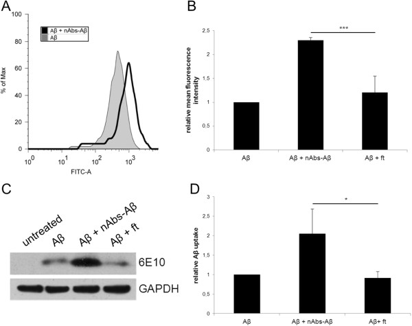
Primary microglial cells isolated from Swiss Webster mouse mesencephalons on embryonic day 13.5 were pretreated with nAbs-Aβ and then treated with Aβ oligomers. After 3 hours, phagocytosis as well as western blot analysis were evaluated to measure the amount of phagocytized Aβ. Cell viability was analyzed using an MTT assay 24 hours after treatment. Pro-inflammatory cytokines in the supernatants were analyzed with ELISAs and then we treated primary neuronal cells with these conditioned microglia supernatants. Twenty-four hours later we did a MTT assay of the treated neurons. We further investigated the effect of a single nAbs-Aβ administration on Tg2576 mice in vivo.
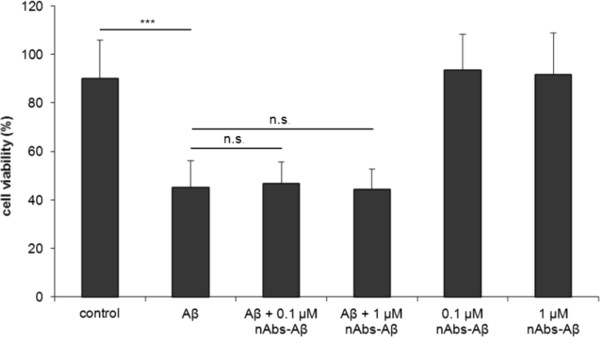
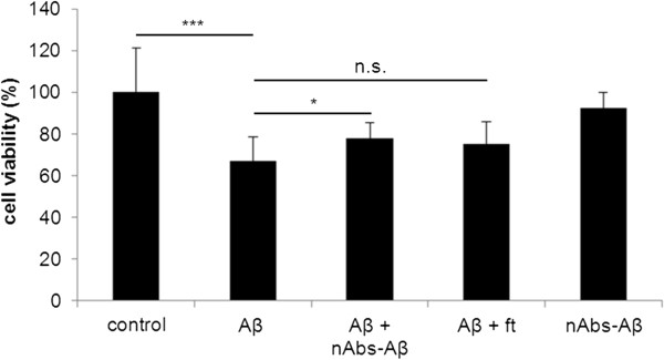
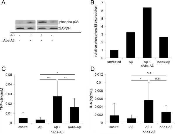
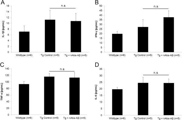
Upon co-administration of Aβ and nAbs-Aβ no change in microglia viability was observed. However, there was an increase in phosphorylated p38 protein level, an increase in the pro-inflammatory cytokines TNF-α and IL-6 and an increase in Aβ uptake by microglial cells. Treatment of primary neurons with conditioned microglia medium led to a 10% improvement in cell viability when nAbs-Aβ were co-administered compared to Aβ-treated cells alone. We were unable to detect changes in cytokine production in brain lysates of Tg2576 mice.
We provide evidence on the mechanism of action of nAbs-Aβ on microglia in vitro. Interestingly, our in vivo data indicate that nAbs-Aβ administration should be considered as a therapeutic strategy in AD, since there is no inflammatory reaction.
天然产生的针对淀粉样蛋白-β(nAbs-Aβ)的自身抗体已被证明对体内转基因阿尔茨海默病(AD)动物和体外原代神经元具有有益作用。但是,对于它们对小胶质细胞的作用知之甚少。我们的目的是研究 nAbs-Aβ 对体外 Aβ 处理的小胶质细胞的影响,包括细胞活力、应激途径、细胞因子产生和吞噬能力,以及这些作用是否可以传递给神经元。
从小鼠胚胎 13.5 天的中脑分离原代小胶质细胞,用 nAbs-Aβ 预处理,然后用 Aβ 寡聚体处理。3 小时后,通过吞噬作用和 Western blot 分析评估吞噬的 Aβ量。处理 24 小时后,通过 MTT 测定分析细胞活力。用 ELISA 分析上清液中的促炎细胞因子,然后用这些条件性小胶质细胞上清液处理原代神经元细胞。24 小时后,用 MTT 测定处理神经元的活力。我们进一步研究了单次 nAbs-Aβ 给药对体内 Tg2576 小鼠的影响。
当 Aβ 和 nAbs-Aβ 共同给药时,小胶质细胞活力没有变化。然而,磷酸化 p38 蛋白水平增加,促炎细胞因子 TNF-α 和 IL-6 增加,小胶质细胞对 Aβ 的摄取增加。用条件性小胶质细胞培养基处理原代神经元,当 nAbs-Aβ 与 Aβ 处理的细胞共同给药时,与单独用 Aβ 处理的细胞相比,细胞活力提高了 10%。我们未能检测到 Tg2576 小鼠脑裂解物中细胞因子产生的变化。
我们提供了 nAbs-Aβ 对体外小胶质细胞作用机制的证据。有趣的是,我们的体内数据表明,nAbs-Aβ 给药应被视为 AD 的一种治疗策略,因为没有炎症反应。