Department of Radiation Oncology, Washington University School of Medicine, Saint Louis, Missouri, USA.
Radiat Oncol. 2013 Jan 17;8:16. doi: 10.1186/1748-717X-8-16.
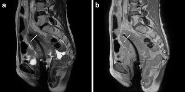
For cervical cancer patients treated with MR-guided high dose rate brachytherapy, the accuracy of radiation delivery depends on accurate localization of both tumors and the applicator, e.g. tandem and ovoid. Standard T2-weighted (T2W) MRI has good tumor-tissue contrast. However, it suffers from poor uterus-tandem contrast, which makes the tandem delineation very challenging. In this study, we evaluated the possibility of using proton density weighted (PDW) MRI to improve the definition of titanium tandems.
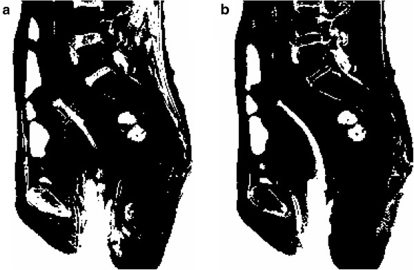
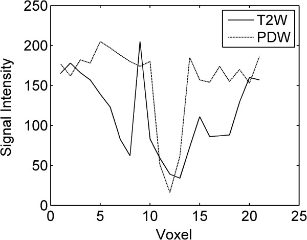
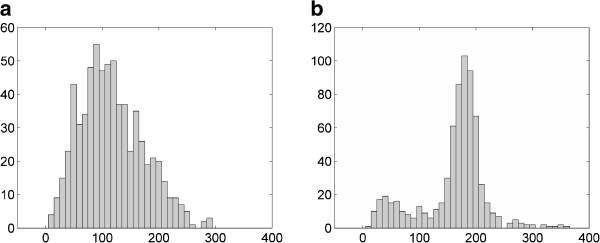
Both T2W and PDW MRI images were obtained from each cervical cancer patient. Imaging parameters were kept the same between the T2W and PDW sequences for each patient except the echo time (90 ms for T2W and 5.5 ms for PDW) and the slice thickness (0.5 cm for T2W and 0.25 cm for PDW). Uterus-tandem contrast was calculated by the equation C=(Su-St)/Su, where Su and St represented the average signal in the uterus and the tandem, respectively. The diameter of the tandem was measured 1.5 cm away from the tip of the tandem. The tandem was segmented by the histogram thresholding technique.
PDW MRI could significantly improve the uterus-tandem contrast compared to T2W MRI (0.42±0.24 for T2W MRI, 0.77±0.14 for PDW MRI, p=0.0002). The average difference between the measured and physical diameters of the tandem was reduced from 0.20±0.15 cm by using T2W MRI to 0.10±0.11 cm by using PDW MRI (p=0.0003). The tandem segmented from the PDW image looked more uniform and complete compared to that from the T2W image.
Compared to the standard T2W MRI, PDW MRI has better uterus-tandem contrast. The information provided by PDW MRI is complementary to those provided by T2W MRI. Therefore, we recommend adding PDW MRI to the simulation protocol to assist tandem delineation process for cervical cancer patients.
对于接受磁共振引导高剂量率近距离放疗的宫颈癌患者,放射治疗的准确性取决于肿瘤和施源器(例如,子宫颈管内照射管和阴道管)的准确定位。标准 T2 加权(T2W)MRI 具有良好的肿瘤组织对比度。然而,它存在子宫-施源器对比度差的问题,这使得施源器勾画极具挑战性。在本研究中,我们评估了使用质子密度加权(PDW)MRI 改善钛制施源器定义的可能性。
每位宫颈癌患者均获得 T2W 和 PDW MRI 图像。对于每位患者,除回波时间(T2W 为 90ms,PDW 为 5.5ms)和切片厚度(T2W 为 0.5cm,PDW 为 0.25cm)外,T2W 和 PDW 序列的成像参数保持一致。子宫-施源器对比度通过公式 C=(Su-St)/Su 计算,其中 Su 和 St 分别代表子宫和施源器的平均信号。在距离施源器尖端 1.5cm 处测量施源器的直径。通过直方图阈值技术对施源器进行分割。
与 T2W MRI 相比,PDW MRI 可显著提高子宫-施源器对比度(T2W MRI 为 0.42±0.24,PDW MRI 为 0.77±0.14,p=0.0002)。使用 T2W MRI 时,测量与物理施源器直径的平均差值为 0.20±0.15cm,而使用 PDW MRI 时,该差值减少至 0.10±0.11cm(p=0.0003)。与 T2W 图像相比,PDW 图像分割的施源器看起来更均匀、完整。
与标准 T2W MRI 相比,PDW MRI 具有更好的子宫-施源器对比度。PDW MRI 提供的信息与 T2W MRI 提供的信息互补。因此,我们建议在模拟方案中添加 PDW MRI,以协助宫颈癌患者的施源器勾画过程。