• 文献检索
  • 文档翻译
  • 深度研究
  • 学术资讯
  • Suppr Zotero 插件Zotero 插件
  • 邀请有礼
  • 套餐&价格
  • 历史记录
应用&插件
Suppr Zotero 插件Zotero 插件浏览器插件Mac 客户端Windows 客户端微信小程序
定价
高级版会员购买积分包购买API积分包
服务
文献检索文档翻译深度研究API 文档MCP 服务
关于我们
关于 Suppr公司介绍联系我们用户协议隐私条款
关注我们

Suppr 超能文献

核心技术专利:CN118964589B侵权必究
粤ICP备2023148730 号-1Suppr @ 2026

文献检索

告别复杂PubMed语法,用中文像聊天一样搜索,搜遍4000万医学文献。AI智能推荐,让科研检索更轻松。

立即免费搜索

文件翻译

保留排版,准确专业,支持PDF/Word/PPT等文件格式,支持 12+语言互译。

免费翻译文档

深度研究

AI帮你快速写综述,25分钟生成高质量综述,智能提取关键信息,辅助科研写作。

立即免费体验

相似文献

1
Single resting hsTnT level predicts abnormal myocardial stress test in acute chest pain patients with normal initial standard troponin.单次静息 hsTnT 水平可预测初始标准肌钙蛋白正常的急性胸痛患者异常心肌应激试验。
JACC Cardiovasc Imaging. 2013 Jan;6(1):72-82. doi: 10.1016/j.jcmg.2012.08.014.
2
Early resting myocardial computed tomography perfusion for the detection of acute coronary syndrome in patients with coronary artery disease.早期静息心肌计算机断层扫描灌注成像用于检测冠心病患者的急性冠状动脉综合征
Circ Cardiovasc Imaging. 2015 Mar;8(3):e002404. doi: 10.1161/CIRCIMAGING.114.002404.
3
High-sensitivity troponin T and C-reactive protein to identify patients without cardiac structural and functional abnormalities as assessed by cardiac CT and SPECT imaging: can biomarkers predict cardiac health?高敏肌钙蛋白 T 和 C 反应蛋白用于识别心脏 CT 和 SPECT 成像评估无心脏结构和功能异常的患者:生物标志物可预测心脏健康吗?
Int J Cardiovasc Imaging. 2013 Apr;29(4):865-73. doi: 10.1007/s10554-012-0164-8. Epub 2013 Jan 1.
4
Gated-SPECT myocardial perfusion imaging and coronary calcium score for evaluation of patients with acute chest pain and a normal or nondiagnostic electrocardiogram.门控单光子发射计算机断层扫描心肌灌注成像和冠状动脉钙化评分用于评估急性胸痛且心电图正常或无诊断意义的患者。
Coron Artery Dis. 2012 Nov;23(7):438-44. doi: 10.1097/MCA.0b013e3283576a13.
5
Combined stress myocardial CT perfusion and coronary CT angiography as a feasible strategy among patients presenting with acute chest pain to the emergency department.对于因急性胸痛到急诊科就诊的患者,联合应激心肌 CT 灌注和冠状动脉 CT 血管造影是一种可行的策略。
J Cardiovasc Comput Tomogr. 2021 Mar-Apr;15(2):129-136. doi: 10.1016/j.jcct.2020.06.195. Epub 2020 Jul 29.
6
hs-Troponin I Followed by CT Angiography Improves Acute Coronary Syndrome Risk Stratification Accuracy and Work-Up in Acute Chest Pain Patients: Results From ROMICAT II Trial.高敏肌钙蛋白I联合CT血管造影可提高急性胸痛患者急性冠脉综合征风险分层的准确性及检查效率:ROMICAT II试验结果
JACC Cardiovasc Imaging. 2015 Nov;8(11):1272-1281. doi: 10.1016/j.jcmg.2015.06.016. Epub 2015 Oct 14.
7
Adenosine stress 64- and 256-row detector computed tomography angiography and perfusion imaging: a pilot study evaluating the transmural extent of perfusion abnormalities to predict atherosclerosis causing myocardial ischemia.腺苷负荷64排和256排探测器计算机断层血管造影及灌注成像:一项评估灌注异常透壁范围以预测引起心肌缺血的动脉粥样硬化的初步研究。
Circ Cardiovasc Imaging. 2009 May;2(3):174-82. doi: 10.1161/CIRCIMAGING.108.813766. Epub 2009 Mar 31.
8
Diagnostic accuracy of combined coronary angiography and adenosine stress myocardial perfusion imaging using 320-detector computed tomography: pilot study.320 层螺旋 CT 冠状动脉造影联合腺苷负荷心肌灌注显像诊断准确性的初步研究。
Eur Radiol. 2013 Jul;23(7):1812-21. doi: 10.1007/s00330-013-2788-z. Epub 2013 Feb 21.
9
Effect of Coronary Atherosclerosis and Myocardial Ischemia on Plasma Levels of High-Sensitivity Troponin T and NT-proBNP in Patients With Stable Angina.冠状动脉粥样硬化和心肌缺血对稳定型心绞痛患者血浆高敏肌钙蛋白T及N末端脑钠肽前体水平的影响
Arterioscler Thromb Vasc Biol. 2016 Apr;36(4):757-64. doi: 10.1161/ATVBAHA.115.306818. Epub 2016 Feb 11.
10
Diagnostic performance of exercise bicycle testing and single-photon emission computed tomography: comparison with 64-slice computed tomography coronary angiography.运动自行车测试和单光子发射计算机断层扫描的诊断性能:与 64 层计算机断层扫描冠状动脉造影的比较。
Int J Cardiovasc Imaging. 2012 Mar;28(3):675-84. doi: 10.1007/s10554-010-9785-y. Epub 2011 Jan 8.

引用本文的文献

1
Value of CT‑derived fractional flow reserve in identifying patients with acute myocardial infarction based on coronary computed tomography angiography.基于冠状动脉计算机断层扫描血管造影术的CT衍生血流储备分数在识别急性心肌梗死患者中的价值。
Exp Ther Med. 2023 Oct 17;26(6):558. doi: 10.3892/etm.2023.12258. eCollection 2023 Dec.
2
Effect of Revascularization on Exercise-Induced Changes in Cardiac and Prothrombotic Biomarkers in Patients with Coronary Artery Disease.冠状动脉疾病患者血运重建对运动诱导的心脏和血栓形成生物标志物变化的影响。
Clin Appl Thromb Hemost. 2022 Jan-Dec;28:10760296221094029. doi: 10.1177/10760296221094029.
3
Myocardial Ischemia and Mobilization of Circulating Progenitor Cells.心肌缺血与循环祖细胞的动员
J Am Heart Assoc. 2018 Feb 20;7(4):e007504. doi: 10.1161/JAHA.117.007504. Epub 2018 Feb 10.
4
Use of High-Sensitivity Cardiac Troponin for the Exclusion of Inducible Myocardial Ischemia: A Cohort Study.应用高敏心肌肌钙蛋白排除可诱导性心肌缺血:一项队列研究。
Ann Intern Med. 2018 Dec 4;169(11):751-760. doi: 10.7326/M18-0670. Epub 2018 Nov 6.
5
Cardiac CT in the Emergency Department: Contrasting Evidence from Registries and Randomized Controlled Trials.急诊科心脏 CT:注册研究和随机对照试验的对比证据。
Curr Cardiol Rep. 2018 Mar 8;20(4):24. doi: 10.1007/s11886-018-0965-z.
6
Stronger correlation with myocardial ischemia of high-sensitivity troponin T than other biomarkers.与其他生物标志物相比,高敏肌钙蛋白 T 与心肌缺血的相关性更强。
J Nucl Cardiol. 2019 Oct;26(5):1674-1683. doi: 10.1007/s12350-018-1199-6. Epub 2018 Jan 29.
7
High-Sensitivity Cardiac Troponin I as a Gatekeeper for Coronary Computed Tomography Angiography and Stress Testing in Patients with Acute Chest Pain.高敏心肌肌钙蛋白I作为急性胸痛患者冠状动脉计算机断层扫描血管造影和负荷试验的守门人
Clin Chem. 2017 Nov;63(11):1724-1733. doi: 10.1373/clinchem.2017.275552. Epub 2017 Sep 18.
8
Cardiac CT angiography for evaluation of acute chest pain.心脏CT血管造影用于评估急性胸痛。
Int J Cardiovasc Imaging. 2016 Jan;32(1):101-12. doi: 10.1007/s10554-015-0763-2. Epub 2015 Sep 5.
9
High-sensitivity cardiac troponin T in stable patients undergoing pharmacological stress testing.接受药物负荷试验的稳定患者的高敏心肌肌钙蛋白T
Clin Cardiol. 2015 May;38(5):293-9. doi: 10.1002/clc.22392. Epub 2015 May 8.
10
Early resting myocardial computed tomography perfusion for the detection of acute coronary syndrome in patients with coronary artery disease.早期静息心肌计算机断层扫描灌注成像用于检测冠心病患者的急性冠状动脉综合征
Circ Cardiovasc Imaging. 2015 Mar;8(3):e002404. doi: 10.1161/CIRCIMAGING.114.002404.

本文引用的文献

1
Serial changes in highly sensitive troponin I assay and early diagnosis of myocardial infarction.高敏肌钙蛋白 I 检测的连续变化与心肌梗死的早期诊断。
JAMA. 2011 Dec 28;306(24):2684-93. doi: 10.1001/jama.2011.1896.
2
Association of troponin T detected with a highly sensitive assay and cardiac structure and mortality risk in the general population.高敏肌钙蛋白 T 检测与普通人群中心脏结构和死亡风险的相关性。
JAMA. 2010 Dec 8;304(22):2503-12. doi: 10.1001/jama.2010.1768.
3
Association of serial measures of cardiac troponin T using a sensitive assay with incident heart failure and cardiovascular mortality in older adults.采用敏感检测方法连续检测心脏肌钙蛋白 T 与老年人心力衰竭和心血管死亡率的关系。
JAMA. 2010 Dec 8;304(22):2494-502. doi: 10.1001/jama.2010.1708. Epub 2010 Nov 15.
4
Determinants of troponin release in patients with stable coronary artery disease: insights from CT angiography characteristics of atherosclerotic plaque.稳定型冠状动脉疾病患者肌钙蛋白释放的决定因素:基于动脉粥样硬化斑块 CT 血管造影特征的研究。
Heart. 2011 May;97(10):823-31. doi: 10.1136/hrt.2010.193201. Epub 2010 Sep 30.
5
Association between high-sensitivity cardiac troponin T levels and the predicted cardiovascular risk in middle-aged men without overt cardiovascular disease.高敏心肌肌钙蛋白 T 水平与无明显心血管疾病的中年男性预测心血管风险的关系。
Am Heart J. 2010 Jun;159(6):972-8. doi: 10.1016/j.ahj.2010.02.036.
6
High-sensitive troponin T: a novel biomarker for prognosis and disease severity in patients with pulmonary arterial hypertension.高敏肌钙蛋白 T:肺动脉高压患者预后和疾病严重程度的新型生物标志物。
Clin Sci (Lond). 2010 Jun 2;119(5):207-13. doi: 10.1042/CS20100014.
7
High-sensitivity troponin T concentrations in acute chest pain patients evaluated with cardiac computed tomography.急性胸痛患者行心脏计算机断层扫描评估的高敏肌钙蛋白 T 浓度。
Circulation. 2010 Mar 16;121(10):1227-34. doi: 10.1161/CIRCULATIONAHA.109.893826. Epub 2010 Mar 1.
8
Analytical validation of a high-sensitivity cardiac troponin T assay.高敏心肌肌钙蛋白 T 检测的分析验证。
Clin Chem. 2010 Feb;56(2):254-61. doi: 10.1373/clinchem.2009.132654. Epub 2009 Dec 3.
9
A sensitive cardiac troponin T assay in stable coronary artery disease.稳定型冠状动脉疾病中的敏感型心肌肌钙蛋白 T 检测。
N Engl J Med. 2009 Dec 24;361(26):2538-47. doi: 10.1056/NEJMoa0805299. Epub 2009 Nov 25.
10
Early diagnosis of myocardial infarction with sensitive cardiac troponin assays.采用敏感心肌肌钙蛋白检测法早期诊断心肌梗死
N Engl J Med. 2009 Aug 27;361(9):858-67. doi: 10.1056/NEJMoa0900428.

单次静息 hsTnT 水平可预测初始标准肌钙蛋白正常的急性胸痛患者异常心肌应激试验。

Single resting hsTnT level predicts abnormal myocardial stress test in acute chest pain patients with normal initial standard troponin.

机构信息

Cardiac MR PET CT Program, Department of Radiology, Massachusetts General Hospital, Harvard Medical School, Boston, Massachusetts 02114, USA.

出版信息

JACC Cardiovasc Imaging. 2013 Jan;6(1):72-82. doi: 10.1016/j.jcmg.2012.08.014.

DOI:10.1016/j.jcmg.2012.08.014
PMID:23328564
原文链接:https://pmc.ncbi.nlm.nih.gov/articles/PMC3734805/
Abstract

OBJECTIVES

The goal of this study was to determine the ability of a single, resting high-sensitivity troponin T (hsTnT) measurement to predict abnormal myocardial perfusion imaging (MPI) in patients presenting with acute chest pain to the emergency department (ED).

BACKGROUND

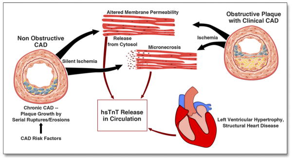
HsTnT assays precisely detect very low levels of troponin T, which may be a surrogate for the presence and extent of myocardial ischemia.

METHODS

We included all patients from the ROMICAT I (Rule Out Myocardial Infarction Using Computer Assisted Tomography) trial, an observational cohort study, who underwent both single-photon emission computed tomography (SPECT)-MPI stress testing and 64-slice computed tomography angiography (CTA) and in whom hsTnT measurements were available. We assessed the discriminatory value of hsTnT for abnormal SPECT-MPI and the association of reversible myocardial ischemia by SPECT-MPI and the extent of coronary atherosclerosis by CTA to hsTnT levels.

RESULTS

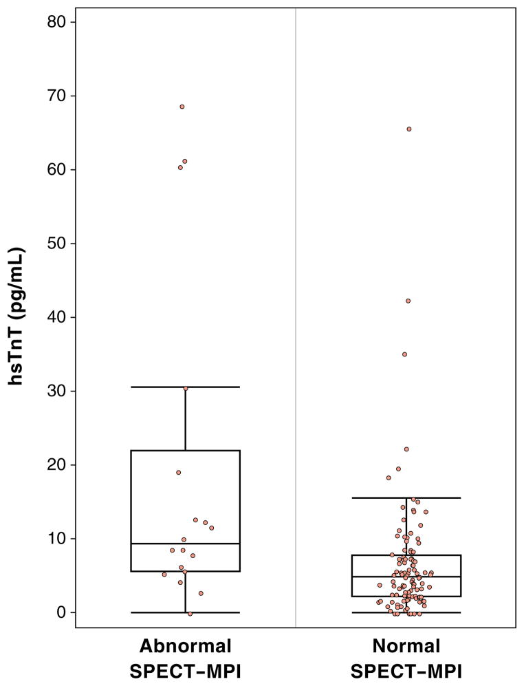
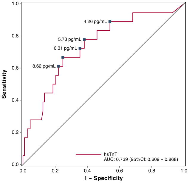
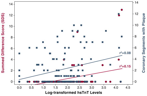
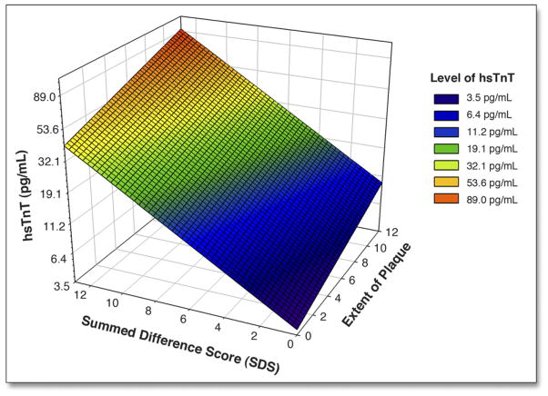
Of the 138 patients (mean age 54 ± 11 years, 46% male), 19 (13.7%) had abnormal SPECT-MPI. Median hsTnT levels were significantly different between patients with normal and abnormal SPECT-MPI (9.41 pg/ml [interquartile range (IQR): 5.73 to 19.20 pg/ml] vs. 4.89 pg/ml [IQR: 2.34 to 7.68 pg/ml], p = 0.001). Sensitivity of 80% and 90% to detect abnormal SPECT-MPI was reached at hsTnT levels as low as 5.73 and 4.26 pg/ml, respectively. Corresponding specificity was 62% and 46%, and negative predictive value was 96% and 96%, respectively. HsTnT levels had good discriminatory ability for prediction of abnormal SPECT-MPI (area under the curve: 0.739, 95% confidence interval: 0.609 to 0.868). Both reversible myocardial ischemia and the extent of coronary atherosclerosis (combined model r(2) = 0.19 with partial of r(2) = 0.12 and r(2) = 0.05, respectively) independently and incrementally predicted the measured hsTnT levels.

CONCLUSIONS

In patients with acute chest pain, myocardial perfusion abnormalities and coronary artery disease are predicted by resting hsTnT levels. Prospective evaluations are warranted to confirm whether resting hsTnT could serve as a powerful triage tool in chest pain patients in the ED before diagnostic testing and improve the effectiveness of patient management.

摘要

目的

本研究旨在确定单次静息高敏肌钙蛋白 T(hsTnT)测量值预测因急性胸痛就诊于急诊科(ED)患者异常心肌灌注成像(MPI)的能力。

背景

hsTnT 检测法可精确检测到极低水平的肌钙蛋白 T,后者可能是心肌缺血存在和严重程度的替代指标。

方法

我们纳入了来自 ROMICAT I(使用计算机辅助断层扫描排除心肌梗死)试验的所有患者,这是一项观察性队列研究,这些患者均同时接受单光子发射计算机断层扫描(SPECT)-MPI 应激试验和 64 层 CT 血管造影(CTA)检查,并且可获得 hsTnT 测量值。我们评估了 hsTnT 对 SPECT-MPI 异常的诊断价值,以及 SPECT-MPI 显示的可逆性心肌缺血和 CTA 显示的冠状动脉粥样硬化程度与 hsTnT 水平之间的相关性。

结果

在 138 例患者(平均年龄 54±11 岁,46%为男性)中,有 19 例(13.7%)患者的 SPECT-MPI 异常。SPECT-MPI 正常和异常患者的 hsTnT 中位数水平差异具有统计学意义(9.41pg/ml [四分位距(IQR):5.73 至 19.20pg/ml] vs. 4.89pg/ml [IQR:2.34 至 7.68pg/ml],p=0.001)。hsTnT 水平分别低至 5.73pg/ml 和 4.26pg/ml 时,其对 SPECT-MPI 异常的检测敏感性分别达到 80%和 90%。相应的特异性分别为 62%和 46%,阴性预测值分别为 96%和 96%。hsTnT 水平对 SPECT-MPI 异常的预测具有良好的判别能力(曲线下面积:0.739,95%置信区间:0.609 至 0.868)。可逆性心肌缺血和冠状动脉粥样硬化程度(联合模型 r²=0.19,偏 r²=0.12,r²=0.05)均独立且呈增量式地预测了测量的 hsTnT 水平。

结论

在因急性胸痛就诊的患者中,静息 hsTnT 水平可预测心肌灌注异常和冠状动脉疾病。需要前瞻性评估来确认静息 hsTnT 是否可作为 ED 胸痛患者在诊断性检查前的强大分诊工具,以改善患者管理的效果。

https://cdn.ncbi.nlm.nih.gov/pmc/blobs/49a1/3734805/b9adcf63f1ba/nihms496248f5.jpg
https://cdn.ncbi.nlm.nih.gov/pmc/blobs/49a1/3734805/9186193d03b1/nihms496248f1.jpg
https://cdn.ncbi.nlm.nih.gov/pmc/blobs/49a1/3734805/c6b56e8f1484/nihms496248f2.jpg
https://cdn.ncbi.nlm.nih.gov/pmc/blobs/49a1/3734805/5a71396fdb98/nihms496248f3.jpg
https://cdn.ncbi.nlm.nih.gov/pmc/blobs/49a1/3734805/d2743b03a812/nihms496248f4.jpg
https://cdn.ncbi.nlm.nih.gov/pmc/blobs/49a1/3734805/b9adcf63f1ba/nihms496248f5.jpg