• 文献检索
  • 文档翻译
  • 深度研究
  • 学术资讯
  • Suppr Zotero 插件Zotero 插件
  • 邀请有礼
  • 套餐&价格
  • 历史记录
应用&插件
Suppr Zotero 插件Zotero 插件浏览器插件Mac 客户端Windows 客户端微信小程序
定价
高级版会员购买积分包购买API积分包
服务
文献检索文档翻译深度研究API 文档MCP 服务
关于我们
关于 Suppr公司介绍联系我们用户协议隐私条款
关注我们

Suppr 超能文献

核心技术专利:CN118964589B侵权必究
粤ICP备2023148730 号-1Suppr @ 2026

文献检索

告别复杂PubMed语法,用中文像聊天一样搜索,搜遍4000万医学文献。AI智能推荐,让科研检索更轻松。

立即免费搜索

文件翻译

保留排版,准确专业,支持PDF/Word/PPT等文件格式,支持 12+语言互译。

免费翻译文档

深度研究

AI帮你快速写综述,25分钟生成高质量综述,智能提取关键信息,辅助科研写作。

立即免费体验

基于模糊C均值算法和形态学运算的子宫肌瘤患者MRI子宫分割与体积测量

Uterine segmentation and volume measurement in uterine fibroid patients' MRI using fuzzy C-mean algorithm and morphological operations.

作者信息

Fallahi Alireza, Pooyan Mohammad, Ghanaati Hossein, Oghabian Mohammad Ali, Khotanlou Hassan, Shakiba Madjid, Jalali Amir Hossein, Firouznia Kavous

机构信息

Department of Biomedical Engineering, Hamedan University of Technology, Hamedan, Iran.

出版信息

Iran J Radiol. 2011 Nov;8(3):150-6. doi: 10.5812/kmp.iranjradiol.17351065.3142. Epub 2011 Nov 25.

DOI:10.5812/kmp.iranjradiol.17351065.3142
PMID:23329932
原文链接:https://pmc.ncbi.nlm.nih.gov/articles/PMC3522330/
Abstract

BACKGROUND

Uterine fibroids are common benign tumors of the female pelvis. Uterine artery embolization (UAE) is an effective treatment of symptomatic uterine fibroids by shrinkage of the size of these tumors. Segmentation of the uterine region is essential for an accurate treatment strategy.

OBJECTIVES

In this paper, we will introduce a new method for uterine segmentation in T1W and enhanced T1W magnetic resonance (MR) images in a group of fibroid patients candidated for UAE in order to make a reliable tool for uterine volumetry.

PATIENTS AND METHODS

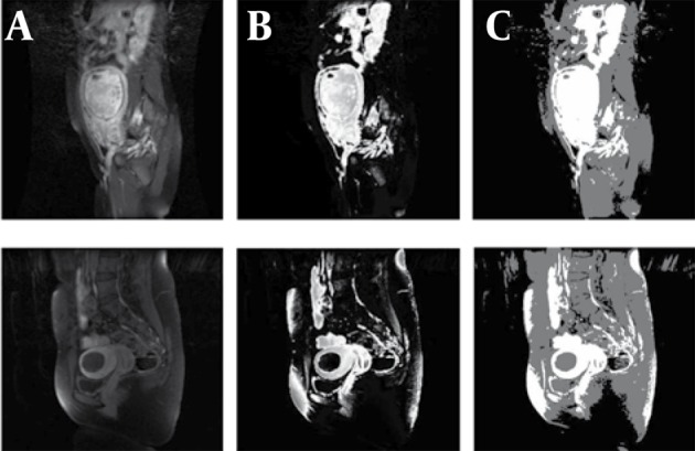
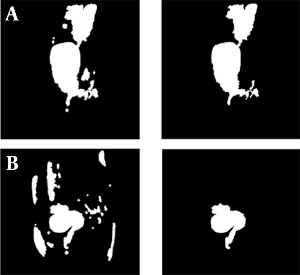
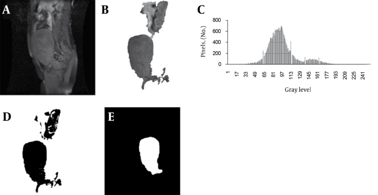
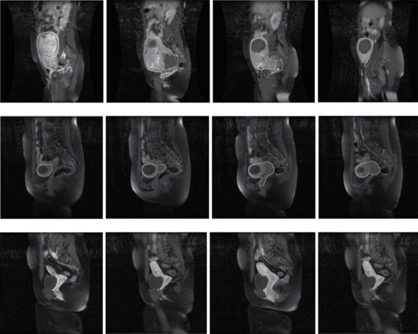
Uterine was initially segmented using Fuzzy C-Mean (FCM) method in T1W-enhanced images and some morphological operations were then applied to refine the initial segmentation. Finally redundant parts were removed by masking the segmented region in T1W-enhanced image over the registered T1W image and using histogram thresholding. This method was evaluated using a dataset with ten patients' images (sagittal, axial and coronal views).

RESULTS

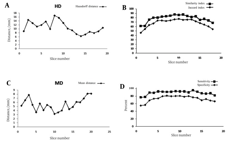
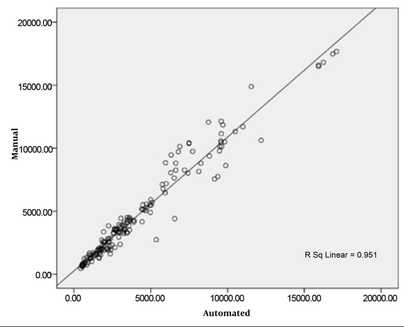
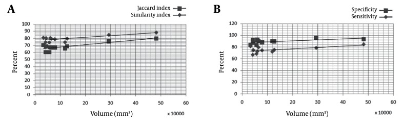
We compared manually segmented images with the output of our system and obtained a mean similarity of 80%, mean sensitivity of 75.32% and a mean specificity of 89.5%. The Pearson correlation coefficient between the areas measured by the manual method and the automated method was 0.99.

CONCLUSIONS

The quantitative results illustrate good performance of this method. By uterine segmentation, fibroids in the uterine may be segmented and their properties may be analyzed.

摘要

背景

子宫肌瘤是女性盆腔常见的良性肿瘤。子宫动脉栓塞术(UAE)是通过缩小这些肿瘤的大小来有效治疗有症状子宫肌瘤的方法。子宫区域的分割对于准确的治疗策略至关重要。

目的

在本文中,我们将介绍一种在一组拟行UAE的子宫肌瘤患者的T1加权和增强T1加权磁共振(MR)图像中进行子宫分割的新方法,以便为子宫容积测量提供可靠工具。

患者和方法

首先在T1加权增强图像中使用模糊C均值(FCM)方法对子宫进行分割,然后应用一些形态学操作来细化初始分割。最后,通过将T1加权增强图像中的分割区域覆盖在配准的T1加权图像上并使用直方图阈值化来去除多余部分。使用包含十名患者图像(矢状面、轴位和冠状面视图)的数据集对该方法进行评估。

结果

我们将手动分割的图像与我们系统的输出进行比较,获得的平均相似度为80%,平均敏感度为75.32%,平均特异度为89.5%。手动测量方法与自动测量方法所测面积之间的Pearson相关系数为0.99。

结论

定量结果表明该方法性能良好。通过子宫分割,可以对子宫内的肌瘤进行分割并分析其特性。

https://cdn.ncbi.nlm.nih.gov/pmc/blobs/be97/3522330/2886daf6558d/iranjradiol-08-150-i003.jpg
https://cdn.ncbi.nlm.nih.gov/pmc/blobs/be97/3522330/dfebe994268e/iranjradiol-08-150-g001.jpg
https://cdn.ncbi.nlm.nih.gov/pmc/blobs/be97/3522330/b7191c3d4cd7/iranjradiol-08-150-g002.jpg
https://cdn.ncbi.nlm.nih.gov/pmc/blobs/be97/3522330/729072fd325c/iranjradiol-08-150-g003.jpg
https://cdn.ncbi.nlm.nih.gov/pmc/blobs/be97/3522330/9dc875d75f9f/iranjradiol-08-150-g004.jpg
https://cdn.ncbi.nlm.nih.gov/pmc/blobs/be97/3522330/492286470c66/iranjradiol-08-150-i001.jpg
https://cdn.ncbi.nlm.nih.gov/pmc/blobs/be97/3522330/d800ee77de1e/iranjradiol-08-150-i002.jpg
https://cdn.ncbi.nlm.nih.gov/pmc/blobs/be97/3522330/2886daf6558d/iranjradiol-08-150-i003.jpg

相似文献

1
Uterine segmentation and volume measurement in uterine fibroid patients' MRI using fuzzy C-mean algorithm and morphological operations.基于模糊C均值算法和形态学运算的子宫肌瘤患者MRI子宫分割与体积测量
Iran J Radiol. 2011 Nov;8(3):150-6. doi: 10.5812/kmp.iranjradiol.17351065.3142. Epub 2011 Nov 25.
2
Uterine artery embolization for symptomatic fibroids in postmenopausal women.绝经后妇女有症状子宫肌瘤的子宫动脉栓塞术。
Clin Imaging. 2016 Jan-Feb;40(1):106-9. doi: 10.1016/j.clinimag.2015.08.010. Epub 2015 Aug 15.
3
A fully automatic 2D segmentation method for uterine fibroid in MRgFUS treatment evaluation.一种用于磁共振引导聚焦超声(MRgFUS)治疗评估中子宫肌瘤的全自动二维分割方法。
Comput Biol Med. 2015 Jul;62:277-92. doi: 10.1016/j.compbiomed.2015.04.030. Epub 2015 Apr 28.
4
Uterine artery embolization for symptomatic fibroids with high signal intensity on T2-weighted MR imaging.磁共振 T2 加权像高信号子宫肌瘤的子宫动脉栓塞术治疗。
Korean J Radiol. 2012 Sep-Oct;13(5):618-24. doi: 10.3348/kjr.2012.13.5.618. Epub 2012 Aug 28.
5
Potential benefit of GnRH-agonist treatment before uterine artery embolization for large fibroids: MRI prediction of fibroid volume reduction.子宫动脉栓塞术前应用 GnRH 激动剂治疗大肌瘤的潜在益处:MRI 预测肌瘤体积缩小。
Acta Radiol. 2022 Oct;63(10):1425-1432. doi: 10.1177/02841851211038802. Epub 2021 Sep 25.
6
Diffusion-weighted MRI with deep learning for visualizing treatment results of MR-guided HIFU ablation of uterine fibroids.基于深度学习的弥散加权磁共振成像用于可视化磁共振引导高强度聚焦超声消融治疗子宫肌瘤的效果。
Eur Radiol. 2023 Jun;33(6):4178-4188. doi: 10.1007/s00330-022-09294-1. Epub 2022 Dec 6.
7
Automated fibroglandular tissue segmentation and volumetric density estimation in breast MRI using an atlas-aided fuzzy C-means method.基于图谱辅助模糊 C 均值法的乳腺 MRI 中纤维腺体组织自动分割及容积密度估测
Med Phys. 2013 Dec;40(12):122302. doi: 10.1118/1.4829496.
8
Combining split-and-merge and multi-seed region growing algorithms for uterine fibroid segmentation in MRgFUS treatments.在磁共振引导聚焦超声(MRgFUS)治疗中,结合分裂合并算法和多种子区域生长算法进行子宫肌瘤分割。
Med Biol Eng Comput. 2016 Jul;54(7):1071-84. doi: 10.1007/s11517-015-1404-6. Epub 2015 Nov 3.
9
Ineffectiveness of magnetic resonance imaging enhancement to predict fibroid volume reduction after uterine artery embolization.磁共振成像增强在预测子宫动脉栓塞术后肌瘤体积缩小方面的无效性。
Proc (Bayl Univ Med Cent). 2017 Jul;30(3):259-261. doi: 10.1080/08998280.2017.11929609.
10
Treatment response assessment of breast masses on dynamic contrast-enhanced magnetic resonance scans using fuzzy c-means clustering and level set segmentation.使用模糊 C 均值聚类和水平集分割评估动态对比增强磁共振扫描中乳腺肿块的治疗反应。
Med Phys. 2009 Nov;36(11):5052-63. doi: 10.1118/1.3238101.

引用本文的文献

1
Artificial intelligence for instance segmentation of MRI: advancing efficiency and safety in laparoscopic myomectomy of broad ligament fibroids.用于磁共振成像实例分割的人工智能:提高阔韧带肌瘤腹腔镜肌瘤切除术的效率和安全性
Front Oncol. 2025 Apr 8;15:1549803. doi: 10.3389/fonc.2025.1549803. eCollection 2025.
2
3D segmentation of uterine fibroids based on deep supervision and an attention gate.基于深度监督和注意力门控的子宫肌瘤三维分割
Front Oncol. 2025 Mar 13;15:1522399. doi: 10.3389/fonc.2025.1522399. eCollection 2025.
3
nnU-Net based segmentation and 3D reconstruction of uterine fibroids with MRI images for HIFU surgery planning.

本文引用的文献

1
Image segmentation via adaptive K-mean clustering and knowledge-based morphological operations with biomedical applications.基于自适应K均值聚类和基于知识的形态学运算的图像分割及其生物医学应用
IEEE Trans Image Process. 1998;7(12):1673-83. doi: 10.1109/83.730379.
2
Extraction of brain tumor from MR images using one-class support vector machine.使用一类支持向量机从磁共振图像中提取脑肿瘤。
Conf Proc IEEE Eng Med Biol Soc. 2005;2005:6411-4. doi: 10.1109/IEMBS.2005.1615965.
3
Role of magnetic resonance imaging in patient selection for uterine artery embolization.
基于 nnU-Net 的 MRI 图像子宫纤维瘤分割与 3D 重建用于 HIFU 手术规划。
BMC Med Imaging. 2024 Sep 6;24(1):233. doi: 10.1186/s12880-024-01385-3.
4
DARU-Net: A dual attention residual U-Net for uterine fibroids segmentation on MRI.DARU-Net:一种用于 MRI 子宫纤维瘤分割的双注意力残差 U-Net。
J Appl Clin Med Phys. 2023 Jun;24(6):e13937. doi: 10.1002/acm2.13937. Epub 2023 Mar 29.
5
Real-time and multimodality image-guided intelligent HIFU therapy for uterine fibroid.实时多模态图像引导的智能高强度聚焦超声治疗子宫肌瘤。
Theranostics. 2020 Mar 26;10(10):4676-4693. doi: 10.7150/thno.42830. eCollection 2020.
6
Evaluation of pre-surgical models for uterine surgery by use of three-dimensional printing and mold casting.通过三维打印和模具铸造对子宫手术术前模型进行评估。
Radiol Phys Technol. 2017 Sep;10(3):279-285. doi: 10.1007/s12194-017-0397-2. Epub 2017 Apr 12.
7
Combining split-and-merge and multi-seed region growing algorithms for uterine fibroid segmentation in MRgFUS treatments.在磁共振引导聚焦超声(MRgFUS)治疗中,结合分裂合并算法和多种子区域生长算法进行子宫肌瘤分割。
Med Biol Eng Comput. 2016 Jul;54(7):1071-84. doi: 10.1007/s11517-015-1404-6. Epub 2015 Nov 3.
8
Uterine artery embolization for treatment of symptomatic fibroids; a single institution experience.子宫动脉栓塞术治疗有症状子宫肌瘤:单机构经验
Hippokratia. 2014 Jul-Sep;18(3):258-61.
9
Uterine artery embolization for treatment of symptomatic fibroids: a review of the evidence.子宫动脉栓塞术治疗有症状子宫肌瘤:证据综述
Iran Red Crescent Med J. 2013 Dec;15(12):e16699. doi: 10.5812/ircmj.16699. Epub 2013 Dec 5.
10
How to start interventional radiology.如何开展介入放射学。
Iran Red Crescent Med J. 2013 Dec;15(12):e16619. doi: 10.5812/ircmj.16619. Epub 2013 Dec 5.
磁共振成像在子宫动脉栓塞术患者选择中的作用。
Acta Radiol. 2006 Dec;47(10):1105-14. doi: 10.1080/02841850600965047.
4
Automatic brain tumor segmentation by subject specific modification of atlas priors.通过对图谱先验进行特定于个体的修改实现脑肿瘤自动分割。
Acad Radiol. 2003 Dec;10(12):1341-8. doi: 10.1016/s1076-6332(03)00506-3.
5
Improved optimization for the robust and accurate linear registration and motion correction of brain images.改进用于脑图像稳健且准确的线性配准和运动校正的优化方法。
Neuroimage. 2002 Oct;17(2):825-41. doi: 10.1016/s1053-8119(02)91132-8.
6
Automatic tumor segmentation using knowledge-based techniques.使用基于知识的技术进行肿瘤自动分割。
IEEE Trans Med Imaging. 1998 Apr;17(2):187-201. doi: 10.1109/42.700731.
7
Changing trends in treatment of leiomyomata uteri.
Curr Opin Obstet Gynecol. 1993 Jun;5(3):301-10.