• 文献检索
  • 文档翻译
  • 深度研究
  • 学术资讯
  • Suppr Zotero 插件Zotero 插件
  • 邀请有礼
  • 套餐&价格
  • 历史记录
应用&插件
Suppr Zotero 插件Zotero 插件浏览器插件Mac 客户端Windows 客户端微信小程序
定价
高级版会员购买积分包购买API积分包
服务
文献检索文档翻译深度研究API 文档MCP 服务
关于我们
关于 Suppr公司介绍联系我们用户协议隐私条款
关注我们

Suppr 超能文献

核心技术专利:CN118964589B侵权必究
粤ICP备2023148730 号-1Suppr @ 2026

文献检索

告别复杂PubMed语法,用中文像聊天一样搜索,搜遍4000万医学文献。AI智能推荐,让科研检索更轻松。

立即免费搜索

文件翻译

保留排版,准确专业,支持PDF/Word/PPT等文件格式,支持 12+语言互译。

免费翻译文档

深度研究

AI帮你快速写综述,25分钟生成高质量综述,智能提取关键信息,辅助科研写作。

立即免费体验

实时多模态图像引导的智能高强度聚焦超声治疗子宫肌瘤。

Real-time and multimodality image-guided intelligent HIFU therapy for uterine fibroid.

机构信息

Department of Biomedical Engineering, School of Medicine, Tsinghua University, Beijing, 100084, China.

National Engineering Research Center of Ultrasound Medicine, Chongqing, 401121, China.

出版信息

Theranostics. 2020 Mar 26;10(10):4676-4693. doi: 10.7150/thno.42830. eCollection 2020.

DOI:10.7150/thno.42830
PMID:32292522
原文链接:https://pmc.ncbi.nlm.nih.gov/articles/PMC7150484/
Abstract

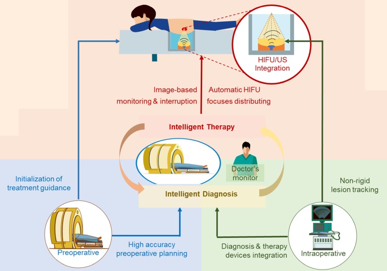
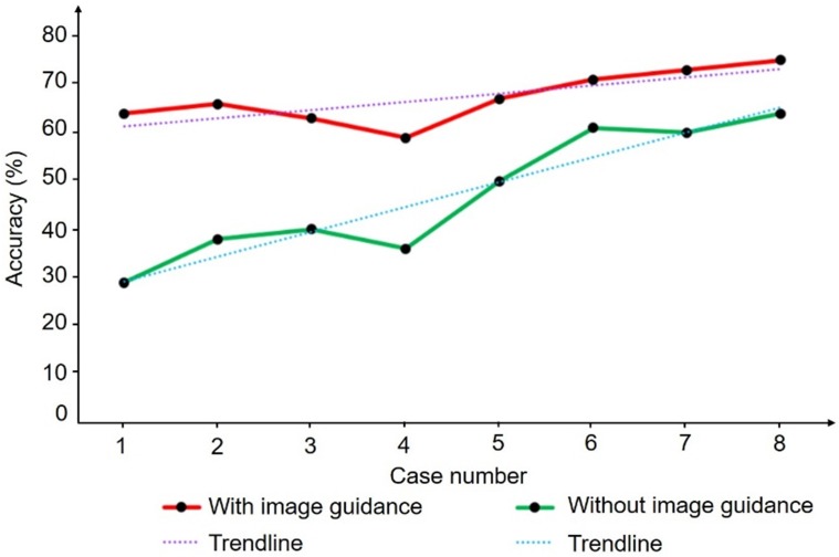
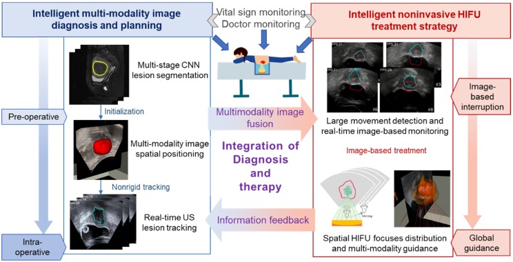
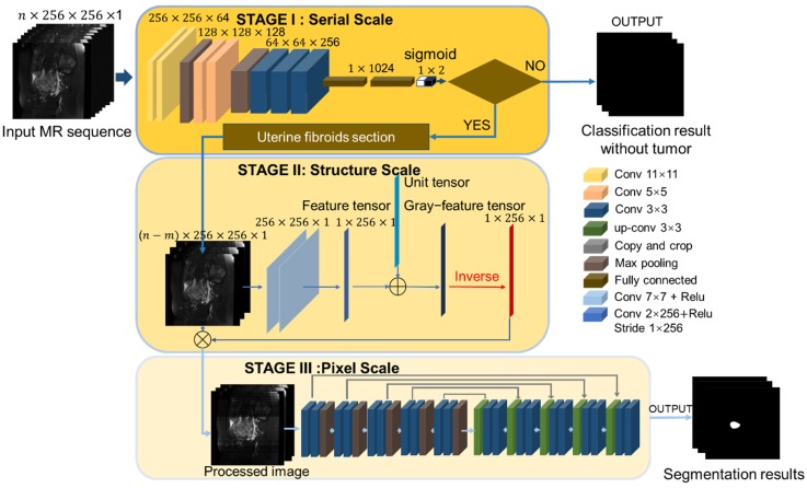
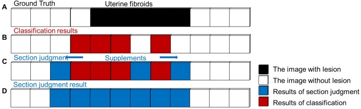
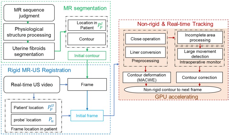
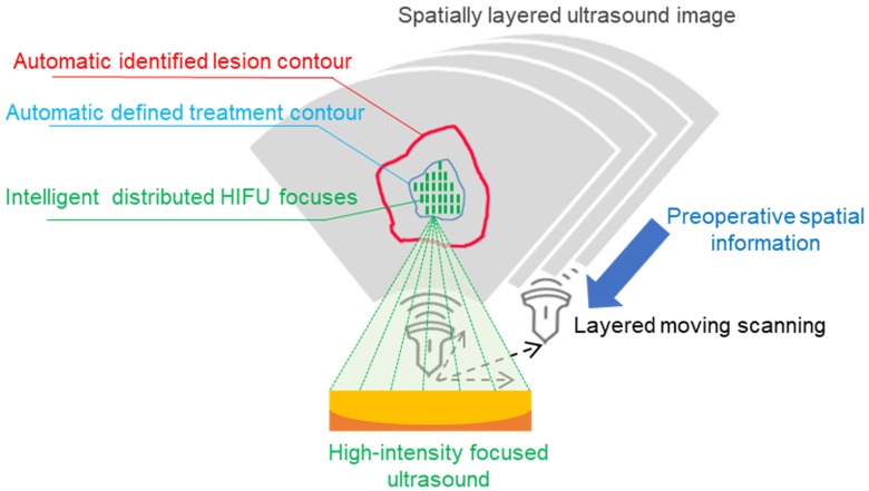
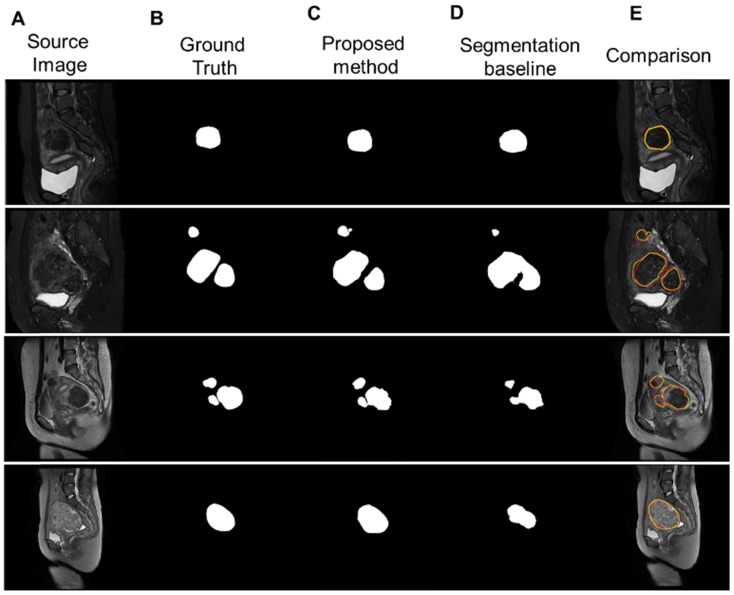
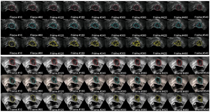
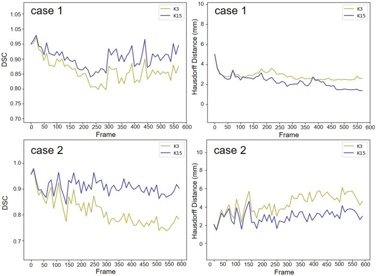
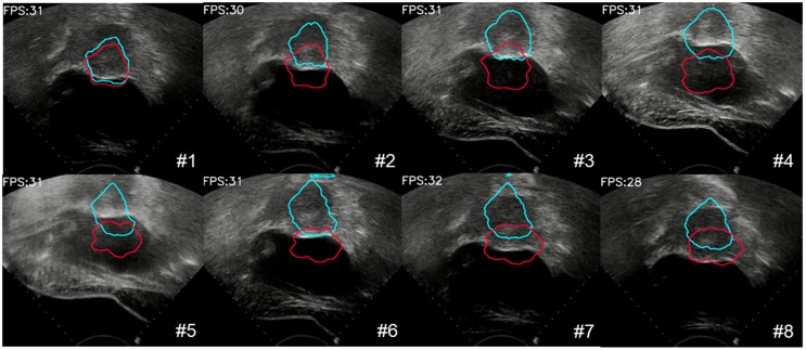
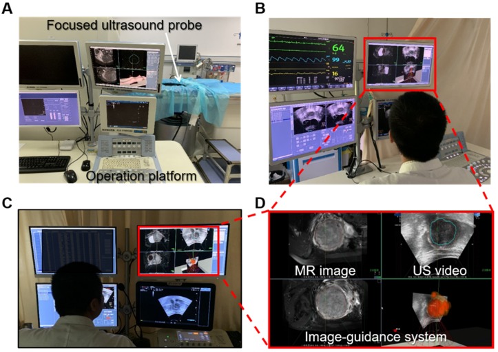
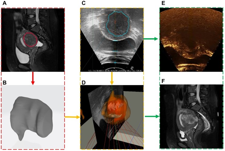
: High-intensity focused ultrasound (HIFU) therapy represents a noninvasive surgical approach to treat uterine fibroids. The operation of HIFU therapy relies on the information provided by medical images. In current HIFU therapy, all operations such as positioning of the lesion in magnetic resonance (MR) and ultrasound (US) images are manually performed by specifically trained doctors. Manual processing is an important limitation of the efficiency of HIFU therapy. In this paper, we aim to provide an automatic and accurate image guidance system, intelligent diagnosis, and treatment strategy for HIFU therapy by combining multimodality information. : In intelligent HIFU therapy, medical information and treatment strategy are automatically processed and generated by a real-time image guidance system. The system comprises a novel multistage deep convolutional neural network for preoperative diagnosis and a nonrigid US lesion tracking procedure for HIFU intraoperative image-assisted treatment. In the process of intelligent therapy, the treatment area is determined from the autogenerated lesion area. Based on the autodetected treatment area, the HIFU foci are distributed automatically according to the treatment strategy. Moreover, an image-based unexpected movement warning and other physiological monitoring are used during the intelligent treatment procedure for safety assurance. : In the experiment, we integrated the intelligent treatment system on a commercial HIFU treatment device, and eight clinical experiments were performed. In the clinical validation, eight randomly selected clinical cases were used to verify the feasibility of the system. The results of the quantitative experiment indicated that our intelligent system met the HIFU clinical tracking accuracy and speed requirements. Moreover, the results of simulated repeated experiments confirmed that the autodistributed HIFU focus reached the level of intermediate clinical doctors. Operations performed by junior- or middle-level operators with the assistance of the proposed system can reach the level of operation performed by senior doctors. Various experiments prove that our proposed intelligent HIFU therapy process is feasible for treating common uterine fibroid cases. : We propose an intelligent HIFU therapy for uterine fibroid which integrates multiple medical information processing procedures. The experiment results demonstrated that the proposed procedures and methods can achieve monitored and automatic HIFU diagnosis and treatment. This research provides a possibility for intelligent and automatic noninvasive therapy for uterine fibroid.

摘要

高强度聚焦超声(HIFU)治疗代表了一种非侵入性的手术方法,可用于治疗子宫肌瘤。HIFU 治疗的操作依赖于医学图像提供的信息。在当前的 HIFU 治疗中,所有操作,如磁共振(MR)和超声(US)图像中病变的定位,都是由经过专门培训的医生手动完成的。手动处理是 HIFU 治疗效率的一个重要限制。在本文中,我们旨在通过结合多模态信息,为 HIFU 治疗提供一种自动、准确的图像引导系统、智能诊断和治疗策略。

在智能 HIFU 治疗中,医疗信息和治疗策略由实时图像引导系统自动处理和生成。该系统包括一个用于术前诊断的新型多级深度卷积神经网络和一个用于 HIFU 术中图像辅助治疗的非刚性 US 病变跟踪程序。在智能治疗过程中,从自动生成的病变区域确定治疗区域。基于自动检测到的治疗区域,根据治疗策略自动分布 HIFU 焦点。此外,在智能治疗过程中使用基于图像的意外运动警告和其他生理监测,以确保安全。

在实验中,我们将智能治疗系统集成到商用 HIFU 治疗设备上,并进行了八项临床实验。在临床验证中,我们使用了 8 个随机选择的临床病例来验证系统的可行性。定量实验的结果表明,我们的智能系统满足 HIFU 临床跟踪精度和速度要求。此外,模拟重复实验的结果证实,自动分布的 HIFU 焦点达到了中级临床医生的水平。在该系统的辅助下,由初级或中级操作人员进行的操作可以达到由高级医生进行的操作水平。各种实验证明,我们提出的智能 HIFU 治疗过程对于治疗常见的子宫肌瘤病例是可行的。

我们提出了一种用于治疗子宫肌瘤的智能 HIFU 治疗方法,该方法集成了多个医疗信息处理程序。实验结果表明,所提出的程序和方法可以实现监控和自动 HIFU 诊断和治疗。这项研究为子宫肌瘤的智能和自动无创治疗提供了一种可能性。

https://cdn.ncbi.nlm.nih.gov/pmc/blobs/fbdf/7150484/c61343104785/thnov10p4676g015.jpg
https://cdn.ncbi.nlm.nih.gov/pmc/blobs/fbdf/7150484/349ab5ac1658/thnov10p4676g001.jpg
https://cdn.ncbi.nlm.nih.gov/pmc/blobs/fbdf/7150484/d012a6408b1c/thnov10p4676g002.jpg
https://cdn.ncbi.nlm.nih.gov/pmc/blobs/fbdf/7150484/77f52e499d38/thnov10p4676g003.jpg
https://cdn.ncbi.nlm.nih.gov/pmc/blobs/fbdf/7150484/860f95b1b98e/thnov10p4676g004.jpg
https://cdn.ncbi.nlm.nih.gov/pmc/blobs/fbdf/7150484/c0a6bcb2185e/thnov10p4676g005.jpg
https://cdn.ncbi.nlm.nih.gov/pmc/blobs/fbdf/7150484/508a0271f1b6/thnov10p4676g006.jpg
https://cdn.ncbi.nlm.nih.gov/pmc/blobs/fbdf/7150484/84f42c45807a/thnov10p4676g007.jpg
https://cdn.ncbi.nlm.nih.gov/pmc/blobs/fbdf/7150484/8251d740a296/thnov10p4676g008.jpg
https://cdn.ncbi.nlm.nih.gov/pmc/blobs/fbdf/7150484/06c6bfea04e8/thnov10p4676g009.jpg
https://cdn.ncbi.nlm.nih.gov/pmc/blobs/fbdf/7150484/32b893fc454c/thnov10p4676g010.jpg
https://cdn.ncbi.nlm.nih.gov/pmc/blobs/fbdf/7150484/3ad30625218f/thnov10p4676g011.jpg
https://cdn.ncbi.nlm.nih.gov/pmc/blobs/fbdf/7150484/16e9661e5ec2/thnov10p4676g012.jpg
https://cdn.ncbi.nlm.nih.gov/pmc/blobs/fbdf/7150484/deed2e51dad4/thnov10p4676g013.jpg
https://cdn.ncbi.nlm.nih.gov/pmc/blobs/fbdf/7150484/ba98cbc05571/thnov10p4676g014.jpg
https://cdn.ncbi.nlm.nih.gov/pmc/blobs/fbdf/7150484/c61343104785/thnov10p4676g015.jpg

相似文献

1
Real-time and multimodality image-guided intelligent HIFU therapy for uterine fibroid.实时多模态图像引导的智能高强度聚焦超声治疗子宫肌瘤。
Theranostics. 2020 Mar 26;10(10):4676-4693. doi: 10.7150/thno.42830. eCollection 2020.
2
Development and validation of a deep learning-based method for automatic measurement of uterus, fibroid, and ablated volume in MRI after MR-HIFU treatment of uterine fibroids.基于深度学习的方法自动测量子宫肌瘤磁共振引导高强度聚焦超声治疗后子宫、肌瘤和消融体积的开发与验证。
Eur J Radiol. 2024 Sep;178:111602. doi: 10.1016/j.ejrad.2024.111602. Epub 2024 Jul 3.
3
Morphological active contour without edge-based model for real-time and non-rigid uterine fibroid tracking in HIFU treatment.用于高强度聚焦超声治疗中实时非刚性子宫肌瘤跟踪的无边缘形态学主动轮廓模型
Healthc Technol Lett. 2019 Nov 26;6(6):172-175. doi: 10.1049/htl.2019.0067. eCollection 2019 Dec.
4
Feasibility of magnetic resonance imaging-guided high intensity focused ultrasound therapy for ablating uterine fibroids in patients with bowel lies anterior to uterus.磁共振引导高强度聚焦超声治疗子宫前位合并肠曲遮挡子宫肌瘤的可行性。
Eur J Radiol. 2010 Feb;73(2):396-403. doi: 10.1016/j.ejrad.2008.11.002. Epub 2008 Dec 23.
5
Portable ultrasound-guided high-intensity focused ultrasound with functions for safe and rapid ablation: prospective clinical trial for uterine fibroids-short-term and long-term results.便携式超声引导高强度聚焦超声治疗,具有安全快速消融功能:子宫肌瘤的前瞻性临床试验——短期和长期结果。
Eur Radiol. 2020 Mar;30(3):1554-1563. doi: 10.1007/s00330-019-06468-2. Epub 2019 Nov 8.
6
Segmentation of uterine fibroid ultrasound images using a dynamic statistical shape model in HIFU therapy.基于动态统计形状模型的超声图像在 HIFU 治疗中的子宫肌瘤分割。
Comput Med Imaging Graph. 2015 Dec;46 Pt 3:302-14. doi: 10.1016/j.compmedimag.2015.07.004. Epub 2015 Aug 6.
7
Multi-scale and shape constrained localized region-based active contour segmentation of uterine fibroid ultrasound images in HIFU therapy.高强度聚焦超声(HIFU)治疗中子宫肌瘤超声图像的多尺度与形状约束局部区域主动轮廓分割
PLoS One. 2014 Jul 25;9(7):e103334. doi: 10.1371/journal.pone.0103334. eCollection 2014.
8
Segmentation of tumor ultrasound image in HIFU therapy based on texture and boundary encoding.基于纹理和边界编码的高强度聚焦超声(HIFU)治疗中肿瘤超声图像分割
Phys Med Biol. 2015 Mar 7;60(5):1807-30. doi: 10.1088/0031-9155/60/5/1807. Epub 2015 Feb 6.
9
Magnetic resonance-high intensity focused ultrasound (MR-HIFU) therapy of symptomatic uterine fibroids with unrestrictive treatment protocols: A systematic review and meta-analysis.磁共振高强度聚焦超声(MR-HIFU)治疗症状性子宫肌瘤的无限制治疗方案:系统评价和荟萃分析。
Eur J Radiol. 2019 Nov;120:108700. doi: 10.1016/j.ejrad.2019.108700. Epub 2019 Oct 15.
10
Automatic segmentation for detecting uterine fibroid regions treated with MR-guided high intensity focused ultrasound (MR-HIFU).用于检测接受磁共振引导高强度聚焦超声(MR-HIFU)治疗的子宫肌瘤区域的自动分割
Med Phys. 2014 Jul;41(7):073502. doi: 10.1118/1.4881319.

引用本文的文献

1
An optimized multi-task contrastive learning framework for HIFU lesion detection and segmentation.一种用于高强度聚焦超声(HIFU)病灶检测与分割的优化多任务对比学习框架。
Sci Rep. 2025 Aug 13;15(1):29666. doi: 10.1038/s41598-025-99783-2.
2
Comparison of the perioperative complications of high intensity focused ultrasound vs. laparoscopic surgery for uterine fibroids: a retrospective study.高强度聚焦超声与腹腔镜手术治疗子宫肌瘤围手术期并发症的比较:一项回顾性研究。
Front Surg. 2025 Jul 17;12:1568000. doi: 10.3389/fsurg.2025.1568000. eCollection 2025.
3
The efficacy and safety of Chinese herbal medicine Shugan Sanjie decoction in the treatment of uterine fibroids: a systematic review and meta-analysis.

本文引用的文献

1
Strategies for Image-Guided Therapy, Surgery, and Drug Delivery Using Photoacoustic Imaging.基于光声成像的影像引导治疗、手术和药物输送策略。
Theranostics. 2019 Feb 27;9(6):1550-1571. doi: 10.7150/thno.32362. eCollection 2019.
2
Non-rigid registration of 3D ultrasound for neurosurgery using automatic feature detection and matching.基于自动特征检测和匹配的神经外科三维超声非刚性配准。
Int J Comput Assist Radiol Surg. 2018 Oct;13(10):1525-1538. doi: 10.1007/s11548-018-1786-7. Epub 2018 Jun 4.
3
3D Imaging and Quantitative Analysis of Vascular Networks: A Comparison of Ultramicroscopy and Micro-Computed Tomography.
中药疏肝散结汤治疗子宫肌瘤的疗效与安全性:一项系统评价与Meta分析
Front Pharmacol. 2025 Apr 17;16:1508015. doi: 10.3389/fphar.2025.1508015. eCollection 2025.
4
Deep learning assisted detection and segmentation of uterine fibroids using multi-orientation magnetic resonance imaging.基于多方位磁共振成像的深度学习辅助子宫肌瘤检测与分割
Abdom Radiol (NY). 2025 Apr 5. doi: 10.1007/s00261-025-04934-8.
5
3D segmentation of uterine fibroids based on deep supervision and an attention gate.基于深度监督和注意力门控的子宫肌瘤三维分割
Front Oncol. 2025 Mar 13;15:1522399. doi: 10.3389/fonc.2025.1522399. eCollection 2025.
6
What Is the Magical Cavitation Bubble: A Holistic Perspective to Trigger Advanced Bubbles, Nano-Sonocatalysts, and Cellular Sonosensitizers.神奇的空化泡是什么:触发高级气泡、纳米声催化剂和细胞声敏剂的整体视角。
BME Front. 2024 Sep 19;5:0067. doi: 10.34133/bmef.0067. eCollection 2024.
7
The Emerging Landscape for Combating Resistance Associated with Energy-Based Therapies via Nanomedicine.通过纳米医学对抗与能量疗法相关的耐药性的新兴领域。
Adv Mater. 2024 Feb;36(5):e2308286. doi: 10.1002/adma.202308286. Epub 2023 Nov 28.
8
Using Clinician-Patient WeChat Group Communication Data to Identify Symptom Burdens in Patients With Uterine Fibroids Under Focused Ultrasound Ablation Surgery Treatment: Qualitative Study.利用医患微信交流群数据识别聚焦超声消融手术治疗子宫肌瘤患者的症状负担:定性研究
JMIR Form Res. 2023 Sep 1;7:e43995. doi: 10.2196/43995.
9
Effects of high-intensity focused ultrasound combined with levonorgestrel-releasing intrauterine system on patients with adenomyosis.高强度聚焦超声联合左炔诺孕酮宫内缓释系统治疗子宫腺肌病的效果。
Sci Rep. 2023 Jun 19;13(1):9903. doi: 10.1038/s41598-023-37096-y.
10
Automated Uterine Fibroids Detection in Ultrasound Images Using Deep Convolutional Neural Networks.使用深度卷积神经网络在超声图像中自动检测子宫肌瘤
Healthcare (Basel). 2023 May 20;11(10):1493. doi: 10.3390/healthcare11101493.
三维成像和血管网络定量分析:超微镜与微计算机断层扫描的比较。
Theranostics. 2018 Mar 7;8(8):2117-2133. doi: 10.7150/thno.22610. eCollection 2018.
4
Automatic Skin Lesion Segmentation Using Deep Fully Convolutional Networks With Jaccard Distance.基于交并比的深度全卷积网络自动皮肤病变分割。
IEEE Trans Med Imaging. 2017 Sep;36(9):1876-1886. doi: 10.1109/TMI.2017.2695227. Epub 2017 Apr 18.
5
Intensity-based 2D 3D registration for lead localization in robot guided deep brain stimulation.基于强度的二维到三维配准在机器人引导的深部脑刺激中用于电极定位
Phys Med Biol. 2017 Mar 21;62(6):2417-2426. doi: 10.1088/1361-6560/aa5ecd. Epub 2017 Feb 7.
6
Ultrasound image based visual servoing for moving target ablation by high intensity focused ultrasound.基于超声图像的运动目标高强度聚焦超声消融的视觉伺服控制。
Int J Med Robot. 2017 Dec;13(4). doi: 10.1002/rcs.1793. Epub 2016 Dec 20.
7
Combining split-and-merge and multi-seed region growing algorithms for uterine fibroid segmentation in MRgFUS treatments.在磁共振引导聚焦超声(MRgFUS)治疗中,结合分裂合并算法和多种子区域生长算法进行子宫肌瘤分割。
Med Biol Eng Comput. 2016 Jul;54(7):1071-84. doi: 10.1007/s11517-015-1404-6. Epub 2015 Nov 3.
8
An efficient algorithm for calculating the exact Hausdorff distance.计算精确 Hausdorff 距离的有效算法。
IEEE Trans Pattern Anal Mach Intell. 2015 Nov;37(11):2153-63. doi: 10.1109/TPAMI.2015.2408351.
9
Three-Dimensional Quantitative Assessment of Uterine Fibroid Response after Uterine Artery Embolization Using Contrast-Enhanced MR Imaging.使用对比增强磁共振成像对子宫动脉栓塞术后子宫肌瘤反应的三维定量评估
J Vasc Interv Radiol. 2015 May;26(5):670-678.e2. doi: 10.1016/j.jvir.2014.11.020. Epub 2015 Jan 28.
10
Using surface markers for MRI guided breast conserving surgery: a feasibility survey.使用表面标志物进行MRI引导下保乳手术:一项可行性调查。
Phys Med Biol. 2014 Apr 7;59(7):1589-605. doi: 10.1088/0031-9155/59/7/1589. Epub 2014 Mar 10.