Thoreson Wallace B, Mercer Aaron J, Cork Karlene M, Szalewski Robert J
Department of Ophthalmology, University of Nebraska Medical Center, Omaha, NE 68198-5840 , USA.
Mol Vis. 2013;19:16-24. Epub 2013 Jan 7.
Efficient and precise release of glutamate from retinal bipolar cells is ensured by the positioning of L-type Ca(2+) channels close to release sites at the base of the synaptic ribbon. We investigated whether Ca(2+) channels at bipolar cell ribbon synapses are fixed in position or capable of moving in the membrane.
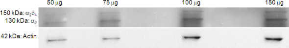
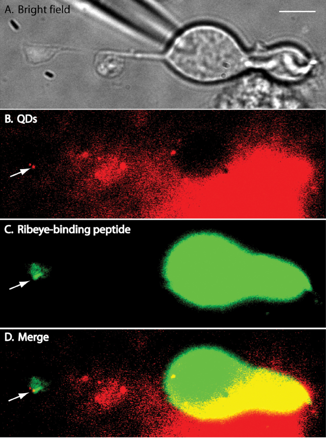
We tracked the movements of individual L-type Ca(2+) channels in bipolar cell terminals after labeling channels with quantum dots (QDs) attached to α(2)δ(4) accessory Ca(2+) channel subunits via intermediary antibodies.
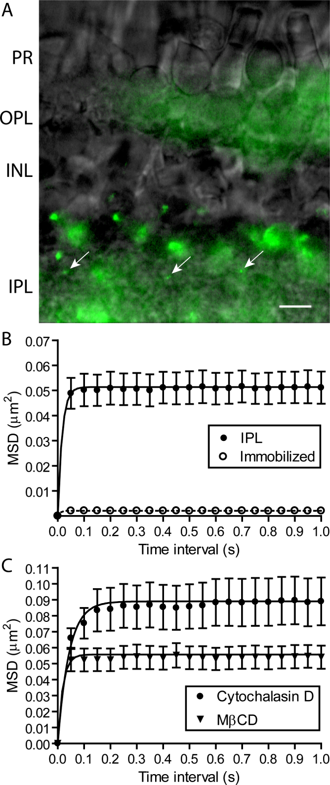
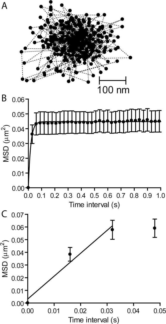
We found that individual Ca(2+) channels moved within a confined domain of 0.13-0.15 μm(2) in bipolar cell terminals, similar to ultrastructural estimates of the surface area of the active zone beneath the ribbon. Disruption of actin expanded the confinement domain indicating that cytoskeletal interactions help to confine channels at the synapse, but the relatively large diffusion coefficients of 0.3-0.45 μm(2)/s suggest that channels are not directly anchored to actin. Unlike photoreceptor synapses, removing membrane cholesterol did not change domain size, indicating that lipid rafts are not required to confine Ca(2+) channels at bipolar cell ribbon synapses.
The ability of Ca(2+) channels to move within the presynaptic active zone suggests that regulating channel mobility may affect release from bipolar cell terminals.
视网膜双极细胞中,L型钙通道定位于突触带基部的释放位点附近,从而确保谷氨酸高效且精确地释放。我们研究了双极细胞带状突触处的钙通道是固定在位还是能够在膜内移动。
我们通过经由中间抗体将量子点(QD)附着于α₂δ₄辅助钙通道亚基来标记通道,之后追踪双极细胞终末中单个L型钙通道的运动。
我们发现,双极细胞终末中单个钙通道在0.13 - 0.15μm²的受限区域内移动,这与突触带下方活性区表面积的超微结构估计值相似。肌动蛋白的破坏扩大了受限区域,表明细胞骨架相互作用有助于将通道限制在突触处,但相对较大的扩散系数为0.3 - 0.45μm²/s,表明通道并非直接锚定在肌动蛋白上。与光感受器突触不同,去除膜胆固醇并未改变区域大小,这表明双极细胞带状突触处限制钙通道并不需要脂筏。
钙通道在突触前活性区内移动的能力表明,调节通道的流动性可能会影响双极细胞终末的释放。