• 文献检索
  • 文档翻译
  • 深度研究
  • 学术资讯
  • Suppr Zotero 插件Zotero 插件
  • 邀请有礼
  • 套餐&价格
  • 历史记录
应用&插件
Suppr Zotero 插件Zotero 插件浏览器插件Mac 客户端Windows 客户端微信小程序
定价
高级版会员购买积分包购买API积分包
服务
文献检索文档翻译深度研究API 文档MCP 服务
关于我们
关于 Suppr公司介绍联系我们用户协议隐私条款
关注我们

Suppr 超能文献

核心技术专利:CN118964589B侵权必究
粤ICP备2023148730 号-1Suppr @ 2026

文献检索

告别复杂PubMed语法,用中文像聊天一样搜索,搜遍4000万医学文献。AI智能推荐,让科研检索更轻松。

立即免费搜索

文件翻译

保留排版,准确专业,支持PDF/Word/PPT等文件格式,支持 12+语言互译。

免费翻译文档

深度研究

AI帮你快速写综述,25分钟生成高质量综述,智能提取关键信息,辅助科研写作。

立即免费体验

胼胝体胶质瘤浸润:DTI 检测到的早期征象。

Glioma infiltration of the corpus callosum: early signs detected by DTI.

机构信息

Neuroradiology, Universitätsmedizin, Georg-August-University Göttingen, Robert-Koch-Str. 40, 37099, Göttingen, Germany.

出版信息

J Neurooncol. 2013 Apr;112(2):217-22. doi: 10.1007/s11060-013-1049-y. Epub 2013 Jan 24.

DOI:10.1007/s11060-013-1049-y
PMID:23344787
原文链接:https://pmc.ncbi.nlm.nih.gov/articles/PMC3607728/
Abstract

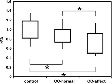
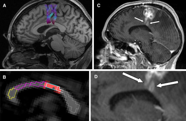
The most frequent primary brain tumors, anaplastic astrocytomas (AA) and glioblastomas (GBM): tend to invasion of the surrounding brain. Histopathological studies found malignant cells in macroscopically unsuspicious brain parenchyma remote from the primary tumor, even affecting the contralateral hemisphere. In early stages, diffuse interneural infiltration with changes of the apparent diffusion coefficient (ADC) and fractional anisotropy (FA) is suspected. The purpose of this study was to investigate the value of DTI as a possible instrument of depicting evidence of tumor invasion into the corpus callosum (CC). Preoperatively, 31 patients with high-grade brain tumors (8 AA and 23 GBM) were examined by MRI at 3 T, applying a high-resolution diffusion tensor imaging (DTI) sequence. ADC- and FA-values were analyzed in the tumor-associated area of the CC as identified by fiber tracking, and were compared to matched healthy controls. In (MR-)morphologically normal appearing CC the ADC values were elevated in the tumor patients (n = 22; 0.978 × 10(-3) mm²/s) compared to matched controls (0.917 × 10(-3) mm²/s, p < 0.05), and the corresponding relative FA was reduced (rFA: 88 %, p < 0.01). The effect was pronounced in case of affection of the CC visible on MRI (n = 9; 0.978 × 10(-3) mm²/s, p < 0.05; rFA: 72 %, p < 0.01). Changes in diffusivity and anisotropy in the CC can be interpreted as an indicator of tumor spread into the contralateral hemisphere not visible on conventional MRI.

摘要

最常见的原发性脑肿瘤,间变性星形细胞瘤(AA)和胶质母细胞瘤(GBM):往往侵袭周围的大脑。组织病理学研究发现,在远离原发肿瘤的宏观上无可疑的脑实质中存在恶性细胞,甚至影响对侧大脑半球。在早期阶段,弥漫性神经内浸润,表观扩散系数(ADC)和各向异性分数(FA)发生变化,故推测弥散张量成像(DTI)可能有助于显示肿瘤向胼胝体(CC)侵袭的证据。术前,对 31 例高级别脑肿瘤患者(8 例 AA 和 23 例 GBM)进行了 3T 磁共振成像(MRI)检查,应用高分辨率弥散张量成像(DTI)序列。通过纤维跟踪术分析肿瘤相关区域的 ADC 和 FA 值,并与匹配的健康对照组进行比较。在(MR)形态上正常的 CC 中,肿瘤患者(n = 22)的 ADC 值升高(0.978×10(-3)mm²/s),与匹配对照组(0.917×10(-3)mm²/s,p < 0.05)相比,相应的相对 FA 降低(rFA:88%,p < 0.01)。在 MRI 可见 CC 受累的情况下(n = 9),这种影响更为明显(0.978×10(-3)mm²/s,p < 0.05;rFA:72%,p < 0.01)。CC 中的弥散性和各向异性变化可解释为肿瘤向对侧半球扩散的指标,在常规 MRI 上不可见。

https://cdn.ncbi.nlm.nih.gov/pmc/blobs/e7f9/7507961/a4d1cbbfa68d/11060_2013_1049_Fig3_HTML.jpg
https://cdn.ncbi.nlm.nih.gov/pmc/blobs/e7f9/7507961/142a56dfed97/11060_2013_1049_Fig1_HTML.jpg
https://cdn.ncbi.nlm.nih.gov/pmc/blobs/e7f9/7507961/986190817a39/11060_2013_1049_Fig2_HTML.jpg
https://cdn.ncbi.nlm.nih.gov/pmc/blobs/e7f9/7507961/a4d1cbbfa68d/11060_2013_1049_Fig3_HTML.jpg
https://cdn.ncbi.nlm.nih.gov/pmc/blobs/e7f9/7507961/142a56dfed97/11060_2013_1049_Fig1_HTML.jpg
https://cdn.ncbi.nlm.nih.gov/pmc/blobs/e7f9/7507961/986190817a39/11060_2013_1049_Fig2_HTML.jpg
https://cdn.ncbi.nlm.nih.gov/pmc/blobs/e7f9/7507961/a4d1cbbfa68d/11060_2013_1049_Fig3_HTML.jpg

相似文献

1
Glioma infiltration of the corpus callosum: early signs detected by DTI.胼胝体胶质瘤浸润:DTI 检测到的早期征象。
J Neurooncol. 2013 Apr;112(2):217-22. doi: 10.1007/s11060-013-1049-y. Epub 2013 Jan 24.
2
Detection of occult neoplastic infiltration in the corpus callosum and prediction of overall survival in patients with glioblastoma using diffusion tensor imaging.使用弥散张量成像检测胼胝体隐匿性肿瘤浸润并预测胶质母细胞瘤患者的总生存期。
Eur J Radiol. 2019 Mar;112:106-111. doi: 10.1016/j.ejrad.2019.01.015. Epub 2019 Jan 15.
3
Abnormalities in the normal appearing white matter of the cerebral hemisphere contralateral to a malignant brain tumor detected by diffusion tensor imaging.通过扩散张量成像检测到的与恶性脑肿瘤对侧大脑半球正常外观白质中的异常情况。
Folia Neuropathol. 2014;52(3):226-33. doi: 10.5114/fn.2014.45563.
4
Diffusion Tensor Imaging Shows Corpus Callosum Differences between High-Grade Gliomas and Metastases.扩散张量成像显示高级别胶质瘤与转移瘤之间胼胝体的差异。
J Neuroimaging. 2018 Mar;28(2):199-205. doi: 10.1111/jon.12478. Epub 2017 Oct 24.
5
Relationships of Language Lateralization with Diffusion Tensor Imaging Metrics of Corpus Callosum, Tumor Grade, and Tumors Distance to Language-Eloquent Areas in Glial Neoplasms.胶质肿瘤中语言偏侧化与胼胝体扩散张量成像指标、肿瘤分级及肿瘤与语言功能区距离的关系
J Comput Assist Tomogr. 2020 Nov/Dec;44(6):956-968. doi: 10.1097/RCT.0000000000001103.
6
Microscopic DTI accurately identifies early glioma cell migration: correlation with multimodal imaging in a new glioma stem cell model.微观扩散张量成像(DTI)准确识别早期胶质瘤细胞迁移:与新的胶质瘤干细胞模型中的多模态成像的相关性
NMR Biomed. 2016 Nov;29(11):1553-1562. doi: 10.1002/nbm.3608. Epub 2016 Sep 15.
7
Corpus callosum injury in patients with diffuse axonal injury: a diffusion tensor imaging study.胼胝体损伤在弥漫性轴索损伤患者中的表现:一项弥散张量成像研究。
NeuroRehabilitation. 2010;26(4):339-45. doi: 10.3233/NRE-2010-0571.
8
Analysis of diffusion tensor imaging metrics for gliomas grading at 3 T.3T 下弥散张量成像指标分析在脑胶质瘤分级中的应用。
Eur J Radiol. 2014 Mar;83(3):e156-65. doi: 10.1016/j.ejrad.2013.12.023. Epub 2014 Jan 4.
9
Value of diffusion tensor imaging in differentiating high-grade from low-grade gliomas.扩散张量成像在鉴别高级别与低级别胶质瘤中的价值。
J Med Assoc Thai. 2013 Jun;96(6):716-21.
10
Quantitative fetal MRI with diffusion tensor imaging in cases with 'short' corpus callosum.应用扩散张量成像的定量胎儿 MRI 在“短”胼胝体病例中的应用。
Ultrasound Obstet Gynecol. 2024 Mar;63(3):385-391. doi: 10.1002/uog.27473. Epub 2024 Feb 7.

引用本文的文献

1
A white matter-centered approach to investigate recurrence pathways in high-grade gliomas: a single-center retrospective study.一种以白质为中心的方法来研究高级别胶质瘤的复发途径:一项单中心回顾性研究。
J Neurooncol. 2025 Jun 3. doi: 10.1007/s11060-025-05050-9.
2
Q-Ball high-resolution fiber tractography: Optimizing corticospinal tract delineation near gliomas and its role in the prediction of postoperative motor deficits- A proof of concept study.Q球高分辨率纤维束成像:优化胶质瘤附近皮质脊髓束的描绘及其在预测术后运动功能缺损中的作用——一项概念验证研究。
Brain Spine. 2024 Nov 14;4:104139. doi: 10.1016/j.bas.2024.104139. eCollection 2024.
3

本文引用的文献

1
Favorable outcome in the elderly cohort treated by concomitant temozolomide radiochemotherapy in a multicentric phase II safety study of 5-ALA.5-ALA 多中心二期安全性研究中,同期替莫唑胺放化疗治疗的老年患者结局良好。
J Neurooncol. 2011 Jun;103(2):361-70. doi: 10.1007/s11060-010-0400-9. Epub 2010 Oct 5.
2
Influence of brain tumors on the MR spectra of healthy brain tissue.脑肿瘤对健康脑组织磁共振波谱的影响。
Magn Reson Med. 2011 Jan;65(1):18-27. doi: 10.1002/mrm.22612.
3
Untreated glioblastoma multiforme: increased myo-inositol and glutamine levels in the contralateral cerebral hemisphere at proton MR spectroscopy.
Single-cell multi-omics sequencing uncovers region-specific plasticity of glioblastoma for complementary therapeutic targeting.
单细胞多组学测序揭示胶质母细胞瘤的区域特异性可塑性,用于互补的治疗靶点。
Sci Adv. 2024 Nov 22;10(47):eadn4306. doi: 10.1126/sciadv.adn4306.
4
Neuroanatomical profiles of cognitive phenotypes in patients with primary brain tumors.原发性脑肿瘤患者认知表型的神经解剖学特征
Neurooncol Adv. 2024 Aug 29;6(1):vdae152. doi: 10.1093/noajnl/vdae152. eCollection 2024 Jan-Dec.
5
Deep intravital brain tumor imaging enabled by tailored three-photon microscopy and analysis.经定制化三光子显微镜和分析实现的脑肿瘤深层活体成像。
Nat Commun. 2024 Sep 10;15(1):7383. doi: 10.1038/s41467-024-51432-4.
6
Diffusion tensor imaging in detecting gliomas sub-regions of infiltration, local and remote recurrences: a systematic review.弥散张量成像检测胶质瘤浸润亚区、局部和远处复发:系统评价。
Neurosurg Rev. 2024 Jul 2;47(1):301. doi: 10.1007/s10143-024-02529-3.
7
Butterfly gliomas: a time for stratified management?蝶骨胶质瘤:分层管理的时代?
Neurosurg Rev. 2023 Sep 4;46(1):223. doi: 10.1007/s10143-023-02126-w.
8
Microstructural Abnormalities in the Contralateral Normal-appearing White Matter of Glioblastoma Patients Evaluated with Advanced Diffusion Imaging.采用先进扩散成像技术评估胶质母细胞瘤患者对侧正常白质的微观结构异常
Magn Reson Med Sci. 2024 Oct 1;23(4):479-486. doi: 10.2463/mrms.mp.2023-0054. Epub 2023 Aug 1.
9
Controllability and Robustness of Functional and Structural Connectomic Networks in Glioma Patients.胶质瘤患者功能和结构连接组网络的可控性与鲁棒性
Cancers (Basel). 2023 May 11;15(10):2714. doi: 10.3390/cancers15102714.
10
The Pathological Activation of Microglia Is Modulated by Sexually Dimorphic Pathways.小胶质细胞的病理性激活受性别二态途径调节。
Int J Mol Sci. 2023 Mar 1;24(5):4739. doi: 10.3390/ijms24054739.
未经治疗的多形性胶质母细胞瘤:质子磁共振波谱显示对侧大脑半球肌醇和谷氨酰胺水平升高。
Radiology. 2009 Dec;253(3):805-12. doi: 10.1148/radiol.2533071654. Epub 2009 Sep 29.
4
Quantitative apparent diffusion coefficients in the characterization of brain tumors and associated peritumoral edema.定量表观扩散系数在脑肿瘤及相关瘤周水肿特征描述中的应用
Acta Radiol. 2009 Jul;50(6):682-9. doi: 10.1080/02841850902933123.
5
Evidence that MR diffusion tensor imaging (tractography) predicts the natural history of regional progression in patients irradiated conformally for primary brain tumors.磁共振扩散张量成像(纤维束成像)可预测原发性脑肿瘤适形放疗患者区域进展自然史的证据。
Int J Radiat Oncol Biol Phys. 2008 Aug 1;71(5):1553-62. doi: 10.1016/j.ijrobp.2008.04.017. Epub 2008 Jun 4.
6
Gender differences and age-related white matter changes of the human brain: a diffusion tensor imaging study.人类大脑的性别差异与年龄相关的白质变化:一项扩散张量成像研究。
Neuroimage. 2008 Jan 15;39(2):566-77. doi: 10.1016/j.neuroimage.2007.09.017. Epub 2007 Sep 20.
7
Detection of tumour infiltration in axonal fibre bundles using diffusion tensor imaging.使用扩散张量成像检测轴突纤维束中的肿瘤浸润
Int J Med Robot. 2005 Sep;1(3):80-6. doi: 10.1002/rcs.31.
8
Increase in glutamate as a sensitive indicator of extracellular matrix integrity in peritumoral edema: a 3.0-tesla proton magnetic resonance spectroscopy study.谷氨酸增加作为瘤周水肿中细胞外基质完整性的敏感指标:一项3.0特斯拉质子磁共振波谱研究
J Neurosurg. 2007 Apr;106(4):609-13. doi: 10.3171/jns.2007.106.4.609.
9
Whole-brain N-acetylaspartate spectroscopy and diffusion tensor imaging in patients with newly diagnosed gliomas: a preliminary study.新诊断胶质瘤患者的全脑 N-乙酰天门冬氨酸光谱分析和弥散张量成像:一项初步研究。
AJNR Am J Neuroradiol. 2006 Nov-Dec;27(10):2137-40.
10
Improved delineation of glioma margins and regions of infiltration with the use of diffusion tensor imaging: an image-guided biopsy study.利用扩散张量成像改善胶质瘤边缘及浸润区域的描绘:一项影像引导活检研究
AJNR Am J Neuroradiol. 2006 Oct;27(9):1969-74.