Department of Psychiatry, Semel Institute for Neuroscience and Human Behavior Center for Cognitive Neuroscience, University of California at Los Angeles, CA 90095, USA.
Bipolar Disord. 2013 Mar;15(2):156-66. doi: 10.1111/bdi.12047. Epub 2013 Jan 24.
We examined resting state functional connectivity in the brain between key emotion regulation regions in bipolar I disorder to delineate differences in coupling from healthy subjects.
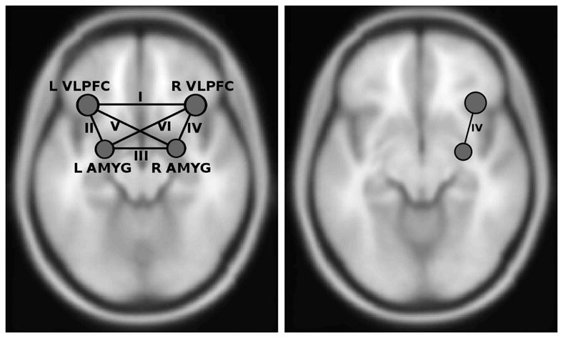
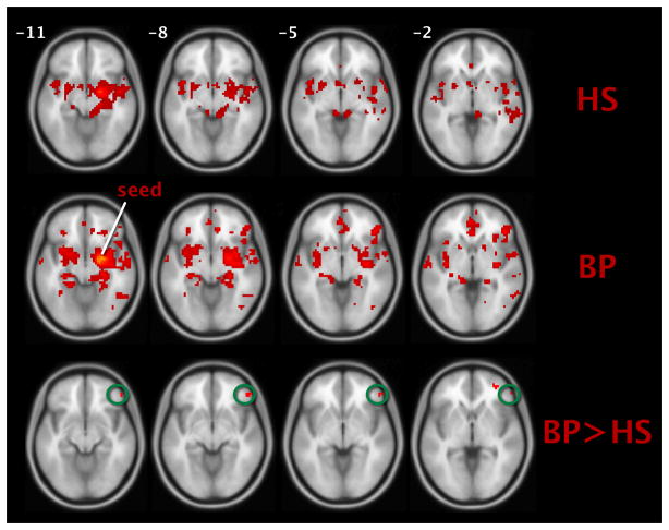
Euthymic subjects with bipolar I disorder (n = 20) and matched healthy subjects (n = 20) participated in a resting state functional magnetic resonance imaging scan. Low-frequency fluctuations in blood oxygen level-dependent (BOLD) signal were correlated in the six connections between four anatomically defined nodes: left and right amygdala and left and right ventrolateral prefrontal cortex (vlPFC). Seed-to-voxel connectivity results were probed for commonly coupled regions. Following this, an identified region was included in a mediation analysis to determine the potential of mediation.
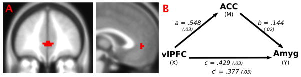
The bipolar I disorder group exhibited significant hyperconnectivity between right amygdala and right vlPFC relative to healthy subjects. The connectivity between these regions in the bipolar I disorder group was partially mediated by activity in the anterior cingulate cortex (ACC).
Greater coupling between right amygdala and right vlPFC and their partial mediation by the ACC were found in bipolar I disorder subjects in remission and in the absence of a psychological task. These findings have implications for a trait-related and clinically important imaging biomarker.
我们研究了双相 I 型障碍患者大脑中关键情绪调节区域之间的静息态功能连接,以描绘与健康受试者的连接差异。
20 名双相 I 型障碍缓解期患者和 20 名匹配的健康受试者参加了静息态功能磁共振成像扫描。在四个解剖定义的节点(左、右杏仁核和左、右腹外侧前额叶皮质)之间的六个连接中,对血氧水平依赖(BOLD)信号的低频波动进行了相关性分析。为了探测共同连接的区域,对种子到体素的连接结果进行了探测。在此之后,将一个确定的区域纳入中介分析,以确定中介的可能性。
与健康受试者相比,双相 I 型障碍组的右杏仁核和右腹外侧前额叶皮质之间存在显著的超连接。在双相 I 型障碍组中,这些区域之间的连接部分被前扣带皮层(ACC)的活动所介导。
在缓解期的双相 I 型障碍患者中,即使在没有心理任务的情况下,也发现右杏仁核和右腹外侧前额叶皮质之间的连接更强,并且其部分被 ACC 所介导。这些发现对与特质相关和临床重要的影像学生物标志物具有影响。