Department of Orthopaedic Surgery, University of Rome Tor Vergata, 81 Oxford Street, Rome 00133, Italy.
BMC Musculoskelet Disord. 2013 Jan 27;14:43. doi: 10.1186/1471-2474-14-43.
Arthroscopic rotator cuff repair has become popular in the last few years because it avoids large skin incisions and deltoid detachment and dysfunction. Earlier arthroscopic single-row (SR) repair methods achieved only partial restoration of the original footprint of the tendons of the rotator cuff, while double-row (DR) repair methods presented many biomechanical advantages and higher rates of tendon-to-bone healing. However, DR repair failed to demonstrate better clinical results than SR repair in clinical trials. MR imaging at 3 Tesla, especially with intra-articular contrast medium (MRA), showed a better diagnostic performance than 1.5 Tesla in the musculoskeletal setting. The objective of this study was to retrospectively evaluate the clinical and 3 Tesla MRA results in two groups of patients operated on for a medium-sized full-thickness rotator cuff tear with two different techniques.
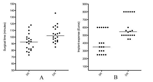
The first group consisted of 20 patients operated on with the SR technique; the second group consisted of 20 patients operated on with the DR technique. All patients were evaluated at a minimum of 3 years after surgery. The primary end point was the re-tear rate at 3 Tesla MRA. The secondary end points were the Constant-Murley Scale (CMS), the Simple Shoulder Test (SST) scores, surgical time and implant expense.
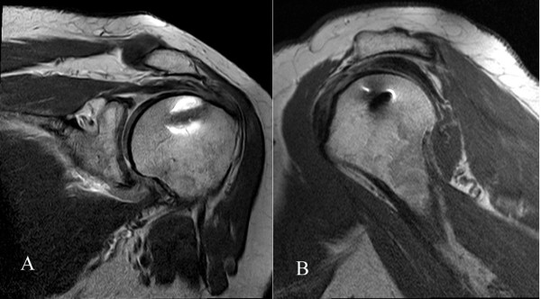
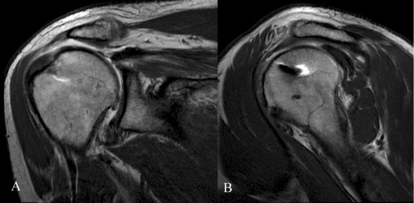
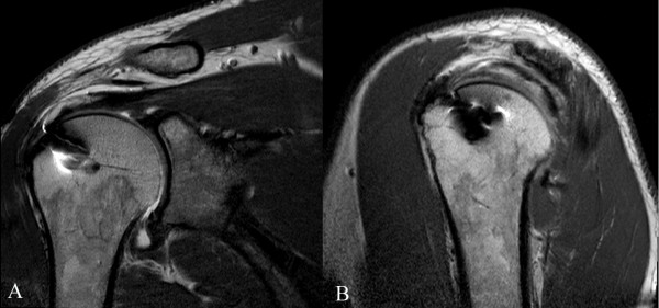
The mean follow-up was 40 months in the SR group and 38.9 months in the DR group. The mean postoperative CMS was 70 in the SR group and 68 in the DR group. The mean SST score was 9.4 in the SR group and 10.1 in the DR group. The re-tear rate was 60% in the SR group and 25% in the DR group. Leakage of the contrast medium was observed in all patients.
To the best of our knowledge, this is the first report on 3 Tesla MRA in the evaluation of two different techniques of rotator cuff repair. DR repair resulted in a statistically significant lower re-tear rate, with longer surgical time and higher implant expense, despite no difference in clinical outcomes. We think that leakage of the contrast medium is due to an incomplete tendon-to-bone sealing, which is not a re-tear. This phenomenon could have important medicolegal implications. Level of evidence III. Treatment study: Case-control study.
关节镜下肩袖修复术在近几年变得流行,因为它避免了大的皮肤切口和三角肌分离及功能障碍。早期的关节镜下单排(SR)修复方法仅实现了肩袖肌腱原始附着点的部分恢复,而双排(DR)修复方法具有许多生物力学优势和更高的肌腱-骨愈合率。然而,DR 修复在临床试验中并未显示出比 SR 修复更好的临床结果。3T 磁共振成像(MR),特别是关节内对比剂(MRA),在肌肉骨骼环境中的诊断性能优于 1.5T。本研究的目的是回顾性评估两组接受不同技术治疗的中等大小全层肩袖撕裂患者的临床和 3T MRA 结果。
第一组包括 20 例采用 SR 技术治疗的患者;第二组包括 20 例采用 DR 技术治疗的患者。所有患者均在术后至少 3 年进行评估。主要终点是 3T MRA 的再撕裂率。次要终点是 Constant-Murley 量表(CMS)、简易肩测试(SST)评分、手术时间和植入物费用。
SR 组的平均随访时间为 40 个月,DR 组为 38.9 个月。SR 组术后 CMS 平均为 70,DR 组为 68。SR 组 SST 评分平均为 9.4,DR 组为 10.1。SR 组再撕裂率为 60%,DR 组为 25%。所有患者均观察到对比剂渗漏。
据我们所知,这是首次报道在评估两种不同肩袖修复技术时使用 3T MRA。DR 修复的再撕裂率明显较低,但手术时间较长,植入物费用较高,尽管临床结果无差异。我们认为对比剂的渗漏是由于肌腱-骨不完全密封,而不是再撕裂。这种现象可能具有重要的医学法律意义。证据水平 III。治疗研究:病例对照研究。