An Tae-Su, Kwon Soon-Il
Department of Ophthalmology, Hallym University Sacred Heart Hospital, Hallym University College of Medicine, Anyang, Korea.
Korean J Ophthalmol. 2013 Feb;27(1):64-7. doi: 10.3341/kjo.2013.27.1.64. Epub 2013 Jan 9.
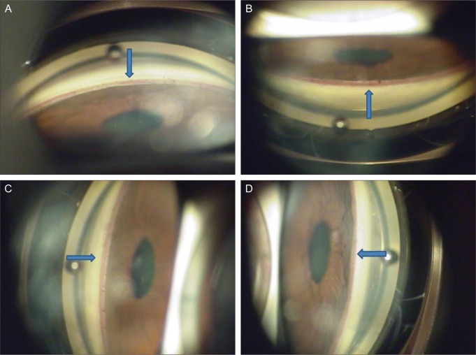
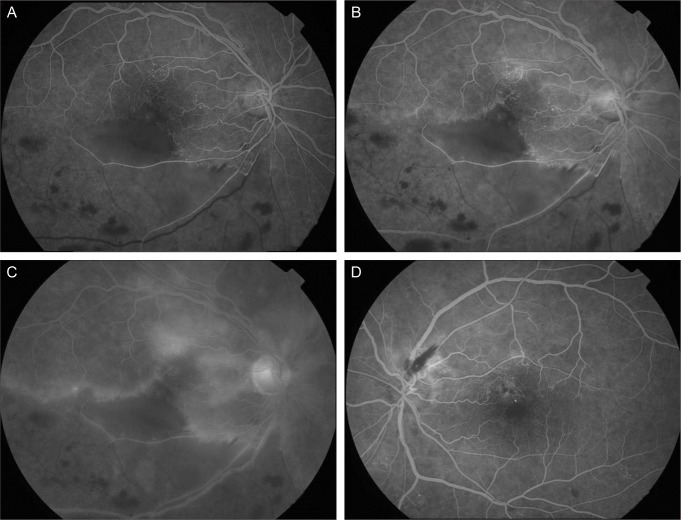
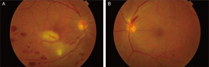
Branch retinal artery occlusion (BRAO) and branch retinal vein occlusion (BRVO) rarely cause neovascular glaucoma (NVG). A 58-year-old woman with hypertension and type 2 diabetic mellitus complained of progressive visual loss in her right eye for the previous 3 months. At initial examination, visual acuity was 20 / 63 in the right eye. Angle neovascularization was observed and the intraocular pressure (IOP) was 30 mmHg in her right eye. Fundus examination and fluorescein angiography showed BRAO combined with BRVO. We immediately injected intravitreal and intracameral bevacizumab in her right eye. The next day, we performed scatter photocoagulation in the nonperfusion area. One month later, visual acuity was 20 / 20 in her right eye and the IOP was 17 mmHg with one topical antiglaucoma agent. The neovascularization had regressed completely. We report a case of unilateral NVG which was caused by BRAO with concomitant BRVO and advise close ophthalmic examination of the iris and angle in BRVO with BRAO.
视网膜分支动脉阻塞(BRAO)和视网膜分支静脉阻塞(BRVO)很少引起新生血管性青光眼(NVG)。一名患有高血压和2型糖尿病的58岁女性,主诉右眼在过去3个月中视力逐渐下降。初诊时,右眼视力为20/63。右眼观察到房角新生血管形成,眼压(IOP)为30 mmHg。眼底检查和荧光素血管造影显示BRAO合并BRVO。我们立即对其右眼进行了玻璃体腔和前房内注射贝伐单抗。第二天,我们对无灌注区进行了散射光凝。1个月后,右眼视力为20/20,使用一种局部抗青光眼药物后眼压为17 mmHg。新生血管已完全消退。我们报告了一例由BRAO合并BRVO引起的单侧NVG病例,并建议对合并BRAO的BRVO患者进行密切的虹膜和房角眼科检查。