Department of Pathology, The Johns Hopkins Hospital, Baltimore, MD 21231, USA.
Hum Pathol. 2013 Jul;44(7):1341-9. doi: 10.1016/j.humpath.2012.11.003. Epub 2013 Jan 31.
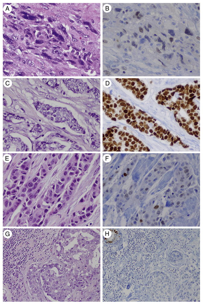
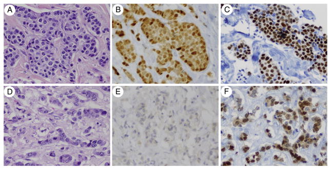
GATA3 plays an integral role in breast luminal cell differentiation and is implicated in breast cancer progression. GATA3 immunohistochemistry is a useful marker of breast cancer; however, its use in specific subtypes is unclear. Here, we evaluate GATA3 expression in 86 invasive ductal carcinomas including triple-negative, Her-2, and luminal subtypes, in addition to 13 metaplastic carcinomas and in 34 fibroepithelial neoplasms. In addition, we report GATA3 expression in matched primary and metastatic breast carcinomas in 30 patients with known estrogen receptor (ER), progesterone receptor (PR), and Her-2 status, including 5 with ER and/or PR loss from primary to metastasis. Tissue microarrays containing 5 to 10 cores per tumor were stained for GATA3, scored as follows: 0 (0-5%), 1+ (6%-25%), 2+ (26%-50%), 3+ (51%-75%), and 4+ (>75%). GATA3 labeling was seen in 67% (66/99) of primary ductal carcinomas including 43% of triple-negative and 54% of metaplastic carcinomas. In contrast, stromal GATA3 labeling was seen in only 1 fibroepithelial neoplasm. GATA3 labeling was seen in 90% (27/30) of primary breast carcinomas in the paired cohort, including 67% of triple-negative carcinomas. GATA3 labeling was overwhelmingly maintained in paired metastases. Notably, GATA3 was maintained in all "luminal loss" metastases, which showed ER and/or PR loss. In conclusion, GATA3 expression is maintained between matched primary and metastatic carcinomas including ER-negative cases. GATA3 can be particularly useful as a marker for metastatic breast carcinoma, especially triple-negative and metaplastic carcinomas, which lack specific markers of mammary origin. Finally, GATA3 labeling may help distinguish metaplastic carcinoma from malignant phyllodes tumors.
GATA3 在乳腺腔细胞分化中起着不可或缺的作用,并与乳腺癌的进展有关。GATA3 免疫组化是乳腺癌的一个有用标志物;然而,其在特定亚型中的应用尚不清楚。在这里,我们评估了 86 例浸润性导管癌(包括三阴性、Her-2 和腔型)、13 例化生癌和 34 例纤维上皮性肿瘤中 GATA3 的表达。此外,我们报告了 30 例已知雌激素受体(ER)、孕激素受体(PR)和 Her-2 状态的原发性和转移性乳腺癌中 GATA3 的表达情况,其中 5 例原发性和转移性乳腺癌中 ER 和/或 PR 丢失。包含每个肿瘤 5 至 10 个核心的组织微阵列用于 GATA3 染色,评分如下:0(0-5%)、1+(6%-25%)、2+(26%-50%)、3+(51%-75%)和 4+(>75%)。在 67%(66/99)的原发性导管癌中观察到 GATA3 标记,包括 43%的三阴性和 54%的化生癌。相比之下,仅在 1 例纤维上皮性肿瘤中观察到基质 GATA3 标记。在配对队列中的 90%(27/30)原发性乳腺癌中观察到 GATA3 标记,包括 67%的三阴性乳腺癌。GATA3 标记在绝大多数配对转移中得到维持。值得注意的是,在所有“腔型丢失”转移中都保留了 GATA3,这些转移均显示 ER 和/或 PR 丢失。总之,GATA3 在匹配的原发性和转移性乳腺癌中保持表达,包括 ER 阴性病例。GATA3 作为转移性乳腺癌的标志物特别有用,尤其是缺乏乳腺来源特异性标志物的三阴性和化生癌。最后,GATA3 标记可能有助于区分化生癌和恶性叶状肿瘤。