Department of Radiation Oncology, Chonnam National University Hwasun Hospital, Chonnam National University Medical School, Gwangju, South Korea.
Department of Biomedical Science, Chonnam National University Graduate School, Gwangju, South Korea.
Front Immunol. 2018 May 2;9:825. doi: 10.3389/fimmu.2018.00825. eCollection 2018.
-expanded natural killer (NK) cells are a potential candidate for cancer immunotherapy based on high cytotoxicity against malignant tumor cells. However, a limited understanding of the migration of activated NK cells toward solid tumors is a critical dilemma in the development of effective and adoptive NK cell-based immunotherapy.
-expanded NK cells from healthy donors were stained with near-infrared fluorophores at different concentrations. NK cell proliferation and cytotoxicity were assessed using a WST-8 assay, while the expression levels of surface molecules were analyzed by flow cytometry. To investigate the biodistribution of NK cells in both normal and tumor-bearing NSG mice, NK cells labeled with ESNF13 were subjected to NIR fluorescence imaging using the Mini-FLARE imaging system. Finally, mice were sacrificed and histopathological tests were performed in resected organs.
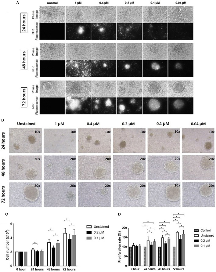
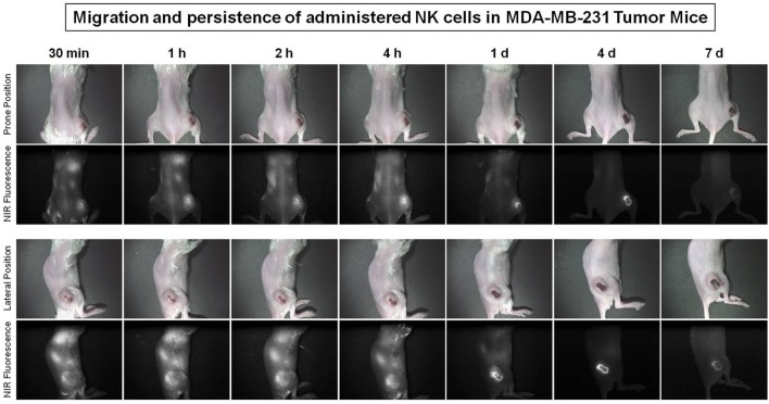
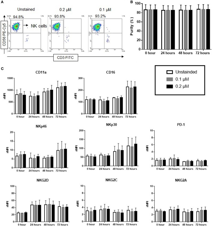
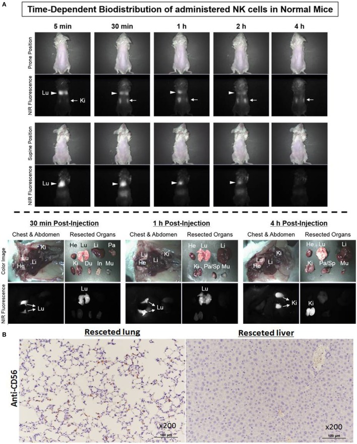
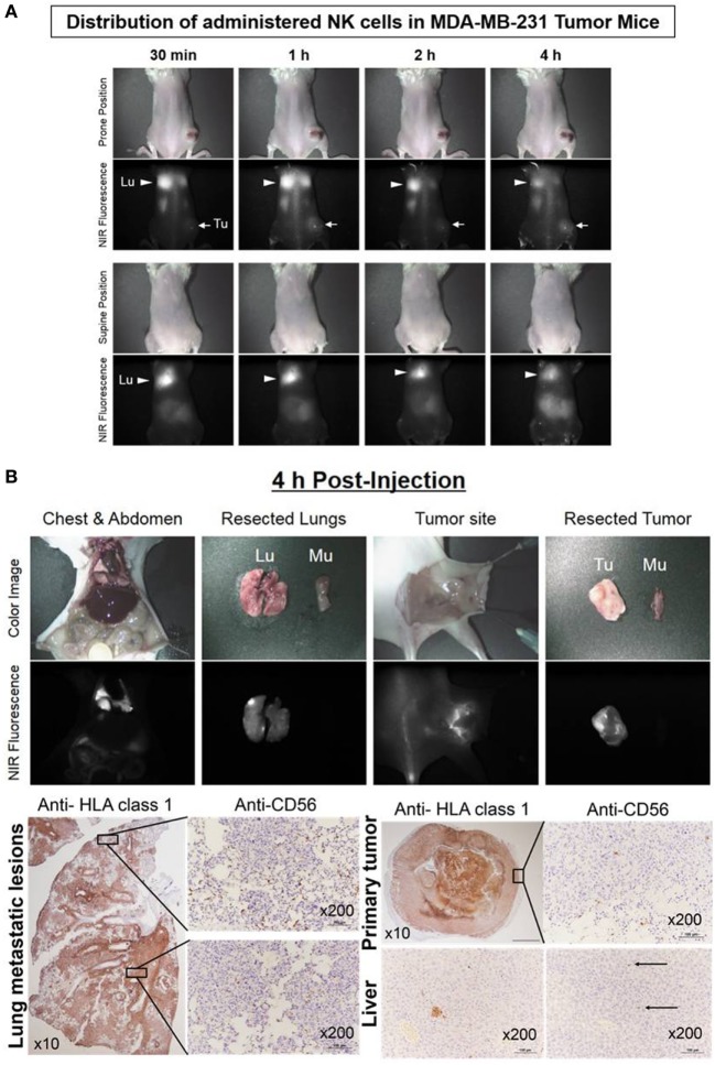
The signal intensity of ESNF-stained NK cells was long-lasting at 72 h using concentrations as low as 0.04 µM. At a low dose range, ESNF13 did not affect NK cell purity, expression levels of surface receptors, or cytotoxic functions against MDA-MB-231 cancer cells. -expanded NK cells labeled with ESNF13 had a 4-h biodistribution in non-tumor-bearing NSG mice that mainly localized to the lungs immediately after injection and then fully migrated to the kidney after 4 h. In an MDA-MB-231 tumor-bearing NSG mice with extensive metastasis in both lungs, the fluorescence signal was dominant in both lungs and steady at 1, 2, and 4 h post-injection. In a early phase of tumor progression, administered NK cell migrated to the lungs and tumor sites within 30 min post-injection, the signal dominated the tumor site after 1 h, and remained steady at 4 h.
Optical imaging with NIR fluorophore ESNF13 is a highly sensitive, applicable, and inexpensive method for the real-time tracking of -expanded NK cells both and . Administered NK cells had different patterns of NK cell distribution and accumulation to the tumor site according to tumor progression in triple-negative breast cancer xenograft models.
用不同浓度的近红外荧光染料对来自健康供体的 NK 细胞进行染色。通过 WST-8 测定法评估 NK 细胞的增殖和细胞毒性,同时通过流式细胞术分析表面分子的表达水平。为了研究 NK 细胞在正常和荷瘤 NSG 小鼠中的分布,用 ESNF13 标记的 NK 细胞进行近红外荧光成像,使用 Mini-FLARE 成像系统。最后,处死小鼠并对切除器官进行组织病理学检查。
用低至 0.04 μM 的浓度,ESNF 染色的 NK 细胞信号强度在 72 小时内保持持久。在低剂量范围内,ESNF13 不会影响 NK 细胞的纯度、表面受体的表达水平或对 MDA-MB-231 癌细胞的细胞毒性作用。用 ESNF13 标记的 - 扩增 NK 细胞在非荷瘤 NSG 小鼠中的分布在 4 小时内,注射后立即主要分布在肺部,然后在 4 小时后完全迁移到肾脏。在 MDA-MB-231 荷瘤 NSG 小鼠中,肺部广泛转移,注射后 1、2 和 4 小时荧光信号均占主导地位。在肿瘤进展的早期阶段,注射后 30 分钟内 NK 细胞迁移到肺部和肿瘤部位,1 小时后信号主导肿瘤部位,4 小时时保持稳定。
用近红外荧光染料 ESNF13 进行光学成像,是一种高度敏感、适用和经济的方法,可实时跟踪 - 扩增的 NK 细胞。根据三阴性乳腺癌异种移植模型中的肿瘤进展,给予的 NK 细胞向肿瘤部位的分布和积累有不同的模式。