Wu Zhenhua, Wang Xinyu, Yang Rong, Liu Yang, Zhao Weiping, Si Jin, Ma Xiaofei, Sun Chao, Liu Yuanyuan, Tan Yong, Liu Wei, Zhang Xin, DI Cuixia, Wang Zhenhua, Zhang Hong, Zhang Zhongxiang
Institute of Modern Physics, Chinese Academy of Sciences, Lanzhou, Gansu 730000; ; Key Laboratory of Heavy Ion Radiation Biology and Medicine of Chinese Academy of Sciences, Lanzhou, Gansu 730000; ; Key Laboratory of Heavy Ion Radiation Medicine of Gansu Province, Lanzhou, Gansu 730000; ; School of Life Science, Lanzhou University, Lanzhou, Gansu 730000;
Exp Ther Med. 2013 Mar;5(3):771-776. doi: 10.3892/etm.2013.881. Epub 2013 Jan 4.
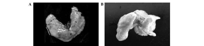
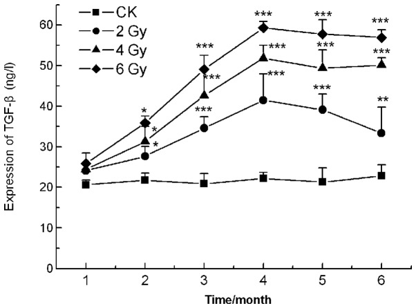
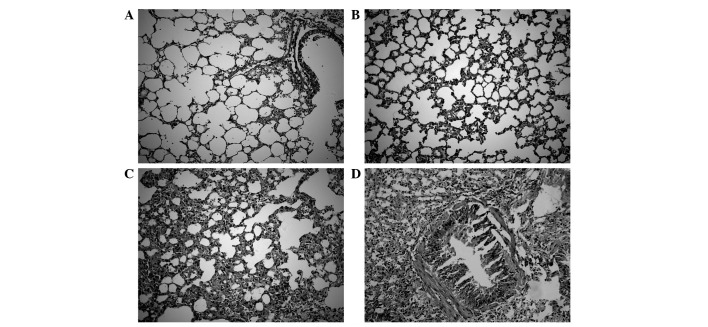
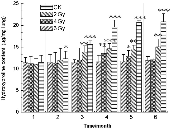
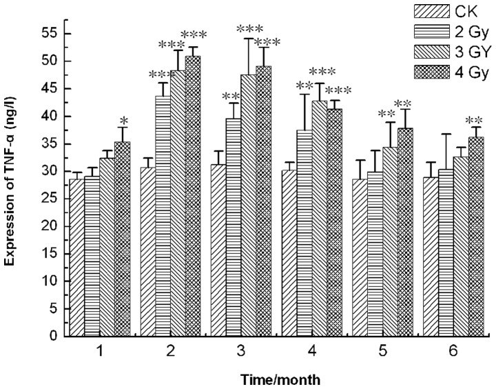
Radiation-induced lung injury is a well-described complication of nuclear accidents, marrow-transplant pretreatment and thoracic radiotherapy. The mechanism is complex and no special therapy for it is available at present. To study radiation pulmonary injury following heavy ion radiotherapy for thoracic tumors, Kunming mice were randomly divided into 4 groups: normal control and 2, 4 and 6 Gy irradiation groups which underwent whole-body exposure to 235 MeV/u (12)C(6+) administered at the Heavy Ion Research Facility in Lanzhou (HIRFL). The pathological changes were observed by hematoxylin and eosin staining and the hydroxyproline (HP) content was assessed by spectrophotometry at months 1, 2, 3, 4, 5 and 6 after radiation exposure. In addition, the expression of tumor necrosis factor (TNF)-α and transforming growth factor (TGF)-β in the lung tissues was measured. The results showed that, compared with the control group, the lung tissue HP content was increased following irradiation but did not statistically significantly change after 4 months in the 4- and 6-Gy-treated groups. However, in the 2-Gy-treated group, the HP content was markedly increased between months 1 and 4 and decreased after month 4. The extent of the lung injury was significantly increased by the higher radiation dosages but was relieved in the 2 Gy group as the time since irradiation increased. The results also revealed that the levels of TNF-α were upregulated and reached a maximum at month 2, but decreased noticeably 2 months later in the experimental groups. The expression of TGF-β increased markedly in month 4 and was altered little in the 4- and 6-Gy-treated groups but decreased sharply in the 2 Gy irradiation group after month 4. These findings suggest that heavy ion radiotherapy for chest tumors causes lung injury to a certain extent, while there is likely to be little injury to lungs treated with <2 Gy, which provides scientific evidence for the use of heavy ion therapy for thoracic tumors.
放射性肺损伤是核事故、骨髓移植预处理及胸部放疗中一种广为人知的并发症。其机制复杂,目前尚无特效治疗方法。为研究胸部肿瘤重离子放疗后的放射性肺损伤,将昆明小鼠随机分为4组:正常对照组以及2、4和6 Gy照射组,这些小鼠在兰州重离子研究装置(HIRFL)接受全身235 MeV/u的碳离子(12C6+)照射。照射后1、2、3、4、5和6个月,通过苏木精-伊红染色观察病理变化,并用分光光度法评估羟脯氨酸(HP)含量。此外,检测肺组织中肿瘤坏死因子(TNF)-α和转化生长因子(TGF)-β的表达。结果显示,与对照组相比,照射后肺组织HP含量升高,但4 Gy和6 Gy治疗组在4个月后无统计学显著变化。然而,2 Gy治疗组在1至4个月间HP含量显著升高,4个月后下降。较高辐射剂量显著增加了肺损伤程度,但2 Gy组随着照射后时间延长损伤减轻。结果还显示,实验组中TNF-α水平上调,在第2个月达到峰值,但2个月后明显下降。TGF-β表达在第4个月显著增加,4 Gy和6 Gy治疗组变化不大,但2 Gy照射组在4个月后急剧下降。这些发现表明,胸部肿瘤重离子放疗会在一定程度上导致肺损伤,而<2 Gy的照射对肺可能损伤较小,这为胸部肿瘤重离子治疗的应用提供了科学依据。