Shioya Sachiko, Masuda Takeshi, Senoo Tadashi, Horimasu Yasushi, Miyamoto Shintaro, Nakashima Taku, Iwamoto Hiroshi, Fujitaka Kazunori, Hamada Hironobu, Hattori Noboru
Department of Molecular and Internal Medicine, Graduate School of Biomedical and Health Sciences, Hiroshima University, Hiroshima 734-8551, Japan.
Department of Respiratory Internal Medicine, Hiroshima University Hospital, Hiroshima 734-8551, Japan.
Exp Ther Med. 2018 Oct;16(4):3070-3076. doi: 10.3892/etm.2018.6550. Epub 2018 Aug 1.
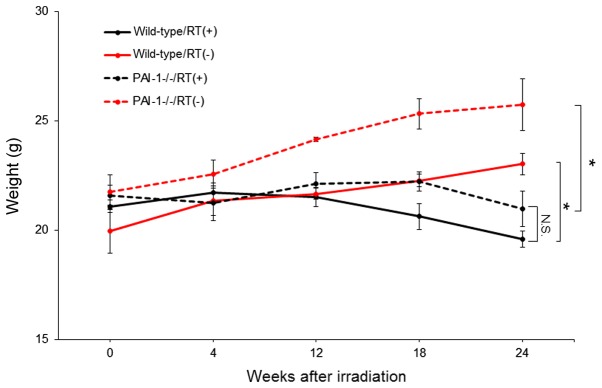
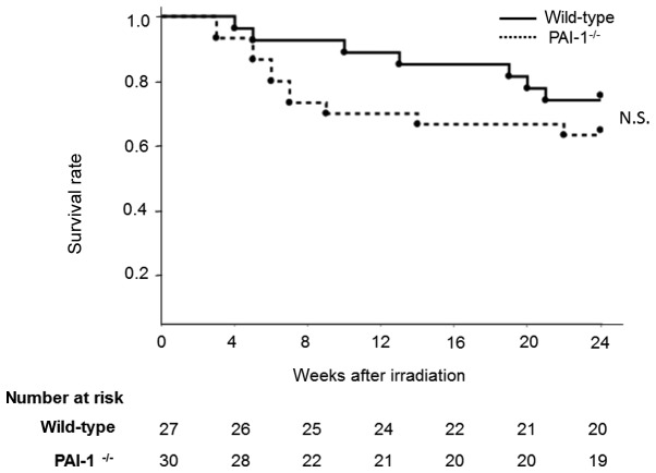
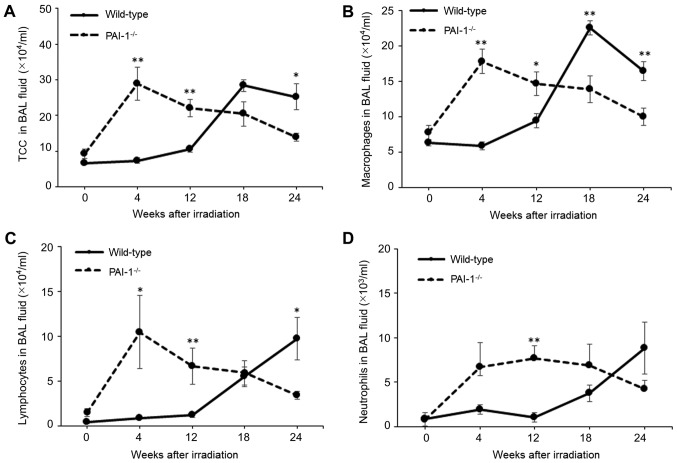
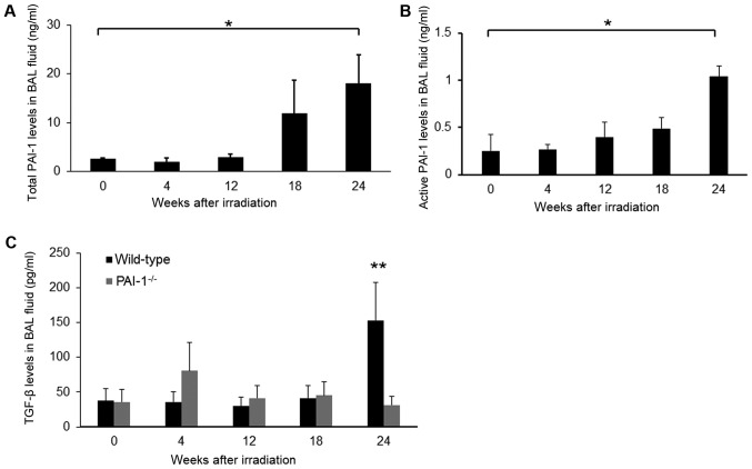
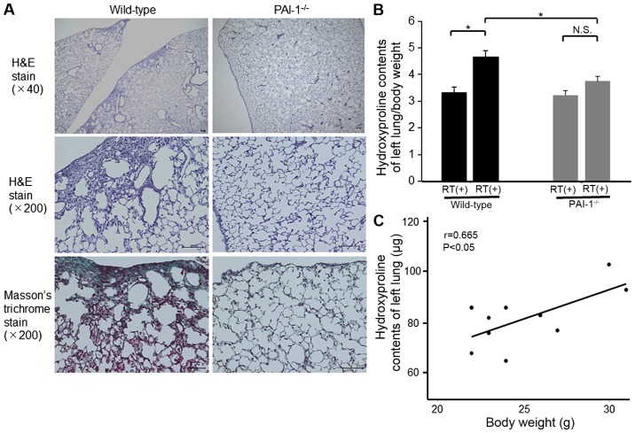
Radiation-induced pulmonary fibrosis is a serious complication. Plasminogen activator inhibitor-1 (PAI-1) has been indicated to be a key factor in the progression of pulmonary fibrosis. In the present study, the effect of PAI-1 deficiency on radiation-induced pulmonary fibrosis was analyzed. Wild-type (WT) and -deficient (PAI-1) mice were treated with thoracic irradiation of 15 Gy to induce pulmonary fibrosis. Analyses of bronchoalveolar lavage (BAL) fluids were performed 0, 4, 12, 18, and 24 weeks after irradiation. The degree of pulmonary fibrosis was assessed according to the histology of lung tissues and hydroxyproline contents. The results demonstrated that the irradiation of WT mice increased PAI-1 expression in the lungs after 18 weeks and established lung fibrosis at 24 weeks. The number of total cells and transforming growth factor-β levels in BAL fluid were significantly lower at 24 weeks after irradiation in PAI-1 mice compared with WT mice. Furthermore, histological examination revealed that the extent of pulmonary fibrosis was attenuated in PAI-1 mice compared with that in WT mice. Hydroxyproline content was also significantly lower in PAI-1 mice compared with WT mice at 24 weeks after irradiation. In conclusion, PAI-1 serves an important role in the development of radiation-induced pulmonary fibrosis and may represent a novel therapeutic target for pulmonary fibrosis.
辐射诱导的肺纤维化是一种严重的并发症。纤溶酶原激活物抑制剂-1(PAI-1)已被证明是肺纤维化进展中的关键因素。在本研究中,分析了PAI-1缺乏对辐射诱导的肺纤维化的影响。对野生型(WT)和PAI-1基因缺陷型小鼠进行15 Gy的胸部照射以诱导肺纤维化。在照射后0、4、12、18和24周对支气管肺泡灌洗(BAL)液进行分析。根据肺组织的组织学和羟脯氨酸含量评估肺纤维化程度。结果表明,WT小鼠在照射18周后肺中PAI-1表达增加,在24周时形成肺纤维化。与WT小鼠相比,PAI-1基因缺陷型小鼠在照射24周后BAL液中的总细胞数和转化生长因子-β水平显著降低。此外,组织学检查显示,与WT小鼠相比,PAI-1基因缺陷型小鼠的肺纤维化程度减轻。照射24周后,PAI-1基因缺陷型小鼠的羟脯氨酸含量也显著低于WT小鼠。总之,PAI-1在辐射诱导的肺纤维化发展中起重要作用,可能是肺纤维化的一个新的治疗靶点。