Department of Health Science, Hamamatsu University School of Medicine,1-20-1 Handa-yama, Higashi-ku, Hamamatsu 431-3192, Japan.
Sci Rep. 2013;3:1255. doi: 10.1038/srep01255. Epub 2013 Feb 13.
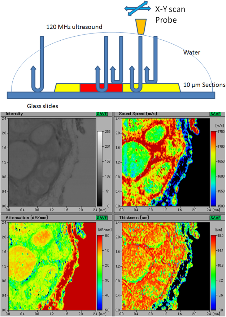
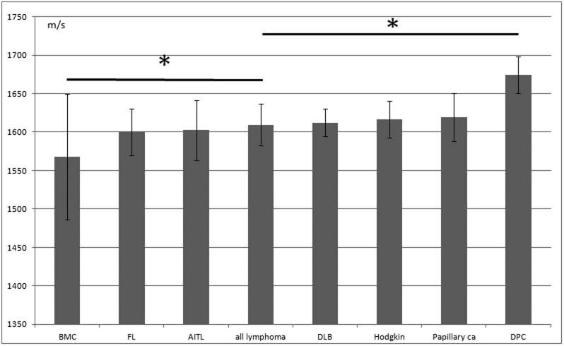
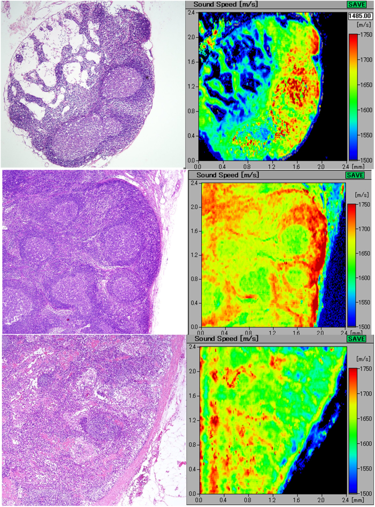
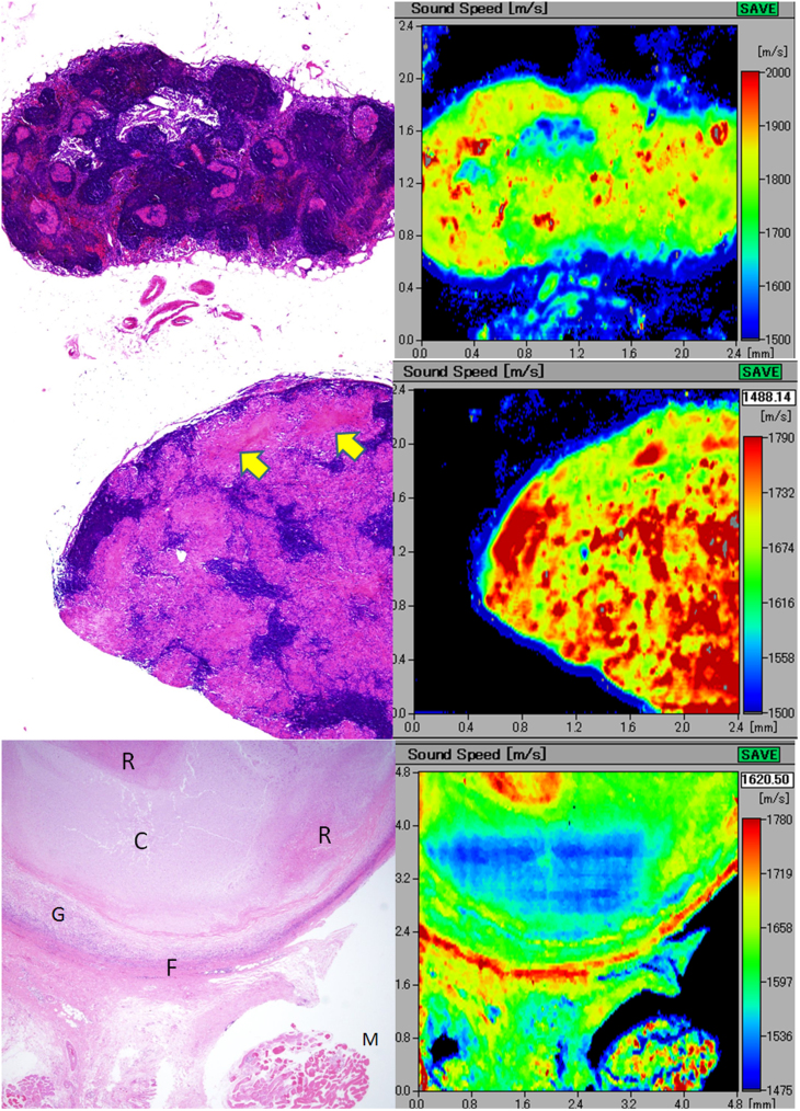
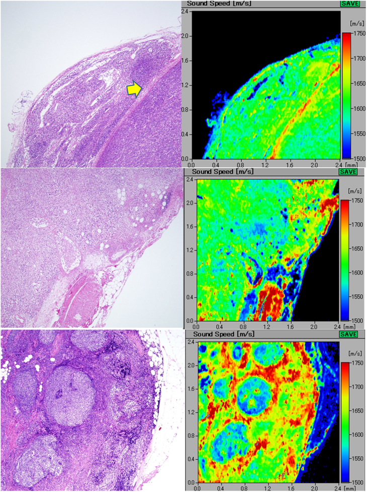
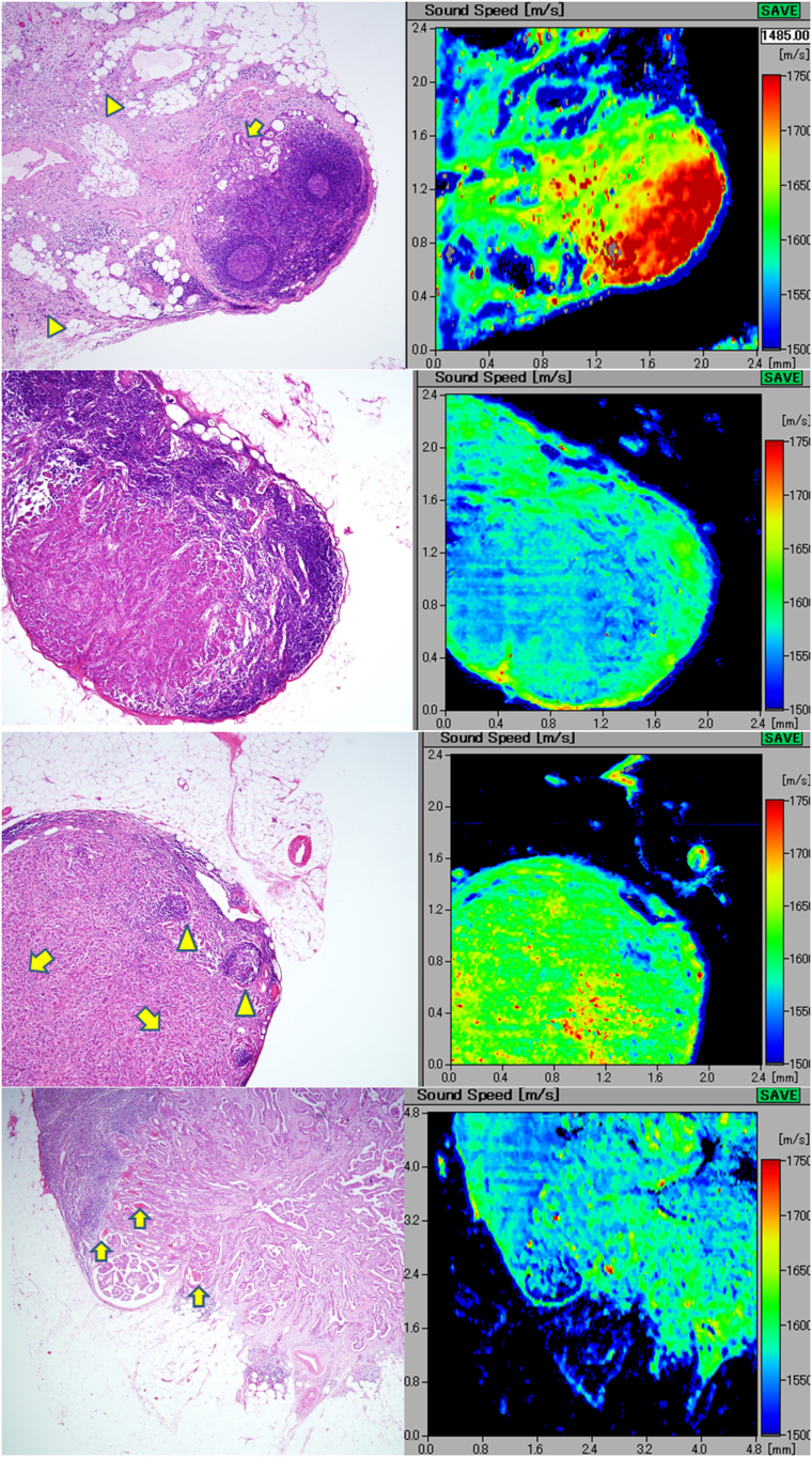
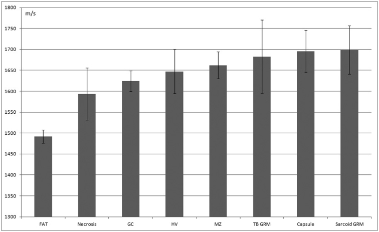
A scanning acoustic microscope (SAM) imaging system calculates and color codes speed of sound (SOS). We evaluated the SAM results for lymph node imaging and compared these results with those of light microscopy (LM). SAM showed normal structures and localized/diffuse lesions of the lymph node. Our results revealed that as a rule, soft areas such as cystic necrosis presented less SOS while harder areas such as coagulative necrosis, granulomas, and fibrosis exhibited greater SOS. SOS increased according to stromal desmoplastic reactions and cellular concentration. In neoplastic lesions, statistically significant differences in SOS were observed among scirrhous carcinomas, lymphomas, and medullary carcinomas. SAM provided the following benefits over LM: (1) images reflected the tissue elasticity of each lesion, (2) digitized SOS data could be statistically comparable, (3) images were acquired in a few minutes without special staining, (4) SAM images and echographic images were comparable for clinical ultrasound imaging study.
扫描声学显微镜(SAM)成像系统计算并对声速(SOS)进行颜色编码。我们评估了 SAM 在淋巴结成像中的结果,并将这些结果与光镜(LM)的结果进行了比较。SAM 显示了淋巴结的正常结构和局部/弥漫性病变。我们的结果表明,通常情况下,囊性坏死等柔软区域的 SOS 较低,而凝固性坏死、肉芽肿和纤维化等较硬区域的 SOS 较高。SOS 随着基质纤维母细胞反应和细胞浓度的增加而增加。在肿瘤病变中,SAM 观察到硬癌、淋巴瘤和髓样癌之间 SOS 存在统计学显著差异。SAM 相对于 LM 具有以下优势:(1)图像反映了每个病变的组织弹性,(2)可对数字化的 SOS 数据进行统计学比较,(3)无需特殊染色即可在几分钟内获取图像,(4)SAM 图像和超声图像可用于临床超声成像研究。