Institute for Medical Immunology, Charité-Universtitätsmedizin Berlin, Berlin, Germany.
ASN Neuro. 2013;5(1):e00110. doi: 10.1042/AN20120081.
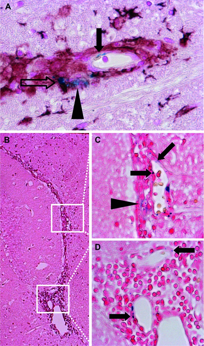
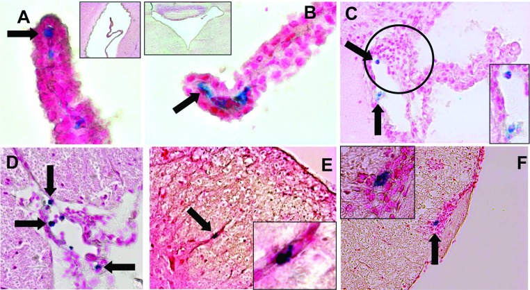
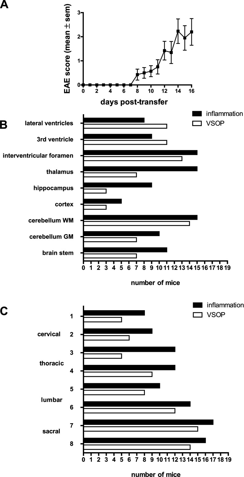
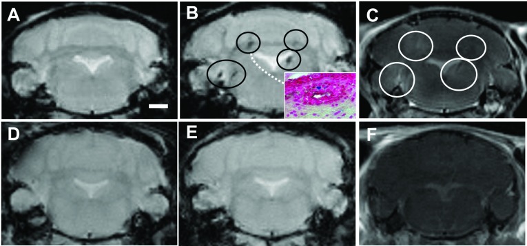
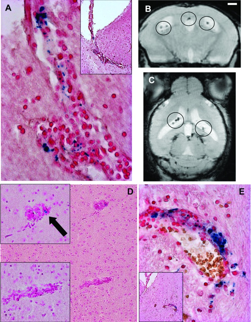
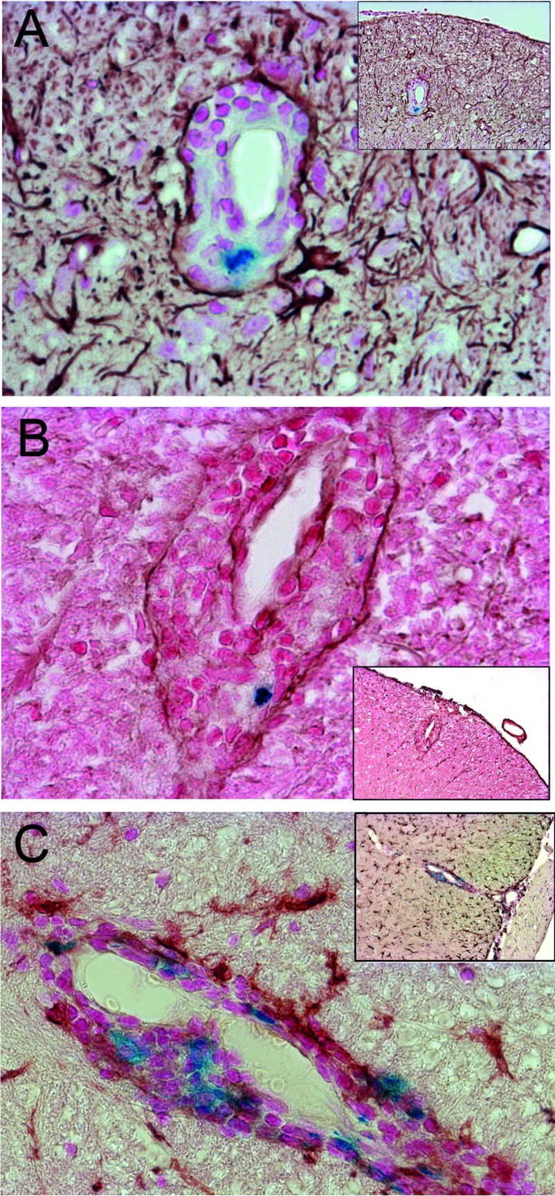
Neuroinflammation during multiple sclerosis involves immune cell infiltration and disruption of the BBB (blood-brain barrier). Both processes can be visualized by MRI (magnetic resonance imaging), in multiple sclerosis patients and in the animal model EAE (experimental autoimmune encephalomyelitis). We previously showed that VSOPs (very small superparamagnetic iron oxide particles) reveal CNS (central nervous system) lesions in EAE which are not detectable by conventional contrast agents in MRI. We hypothesized that VSOP may help detect early, subtle inflammatory events that would otherwise remain imperceptible. To investigate the capacity of VSOP to reveal early events in CNS inflammation, we induced EAE in SJL mice using encephalitogenic T-cells, and administered VSOP prior to onset of clinical symptoms. In parallel, we administered VSOP to mice at peak disease, and to unmanipulated controls. We examined the distribution of VSOP in the CNS by MRI and histology. Prior to disease onset, in asymptomatic mice, VSOP accumulated in the choroid plexus and in spinal cord meninges in the absence of overt inflammation. However, VSOP was undetectable in the CNS of non-immunized control mice. At peak disease, VSOP was broadly distributed; we observed particles in perivascular inflammatory lesions with apparently preserved glia limitans. Moreover, at peak disease, VSOP was prominent in the choroid plexus and was seen in elongated endothelial structures, co-localized with phagocytes, and diffusely disseminated in the parenchyma, suggesting multiple entry mechanisms of VSOP into the CNS. Thus, using VSOP we were able to discriminate between inflammatory events occurring in established EAE and, importantly, we identified CNS alterations that appear to precede immune cell infiltration and clinical onset.
多发性硬化症的神经炎症涉及免疫细胞浸润和血脑屏障 (BBB) 的破坏。这两个过程都可以通过 MRI(磁共振成像)在多发性硬化症患者和实验性自身免疫性脑脊髓炎 (EAE) 动物模型中观察到。我们之前表明,VSOPs(超小超顺磁性氧化铁颗粒)可以揭示 EAE 中的中枢神经系统 (CNS) 病变,而 MRI 中的常规对比剂无法检测到这些病变。我们假设 VSOP 可能有助于检测早期细微的炎症事件,否则这些事件将无法察觉。为了研究 VSOP 揭示中枢神经系统炎症早期事件的能力,我们使用致脑炎 T 细胞在 SJL 小鼠中诱导 EAE,并在出现临床症状之前给予 VSOP。平行地,我们在疾病高峰期给予 VSOP 并给予未处理的对照小鼠。我们通过 MRI 和组织学检查了 VSOP 在中枢神经系统中的分布。在疾病发作前,无症状小鼠中,VSOP 在脉络丛和脊髓脑膜中积累,而没有明显的炎症。然而,未免疫的对照小鼠的中枢神经系统中无法检测到 VSOP。在疾病高峰期,VSOP 广泛分布;我们观察到血管周围炎症病变中有颗粒,胶质界似乎完好无损。此外,在疾病高峰期,VSOP 在脉络丛中很明显,并且在与吞噬细胞共定位的伸长的内皮结构中可见,并在实质中弥散分布,表明 VSOP 进入中枢神经系统的多种进入机制。因此,我们使用 VSOP 能够区分在既定 EAE 中发生的炎症事件,并且重要的是,我们确定了似乎先于免疫细胞浸润和临床发作的中枢神经系统改变。