Silva Rafaela Vieira, Morr Anna S, Mueller Susanne, Koch Stefan Paul, Boehm-Sturm Philipp, Rodriguez-Sillke Yasmina, Kunkel Désirée, Tzschätzsch Heiko, Kühl Anja A, Schnorr Jörg, Taupitz Matthias, Sack Ingolf, Infante-Duarte Carmen
Charité - Universitätsmedizin Berlin, Corporate Member of Freie Universität Berlin and Humboldt-Universität zu Berlin, Institute of Medical Immunology, Berlin, Germany.
Charité - Universitätsmedizin Berlin, Einstein Center for Neurosciences Berlin, Berlin, Germany.
Front Neurosci. 2021 Aug 23;15:701308. doi: 10.3389/fnins.2021.701308. eCollection 2021.
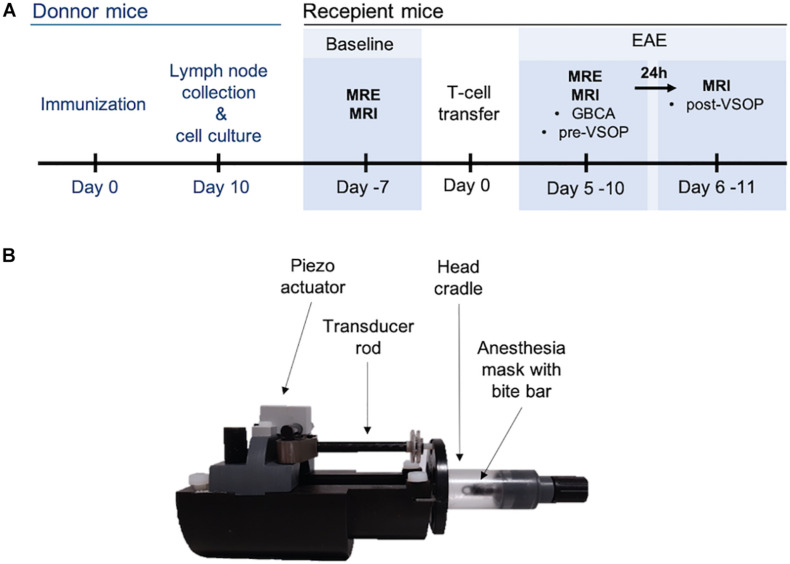
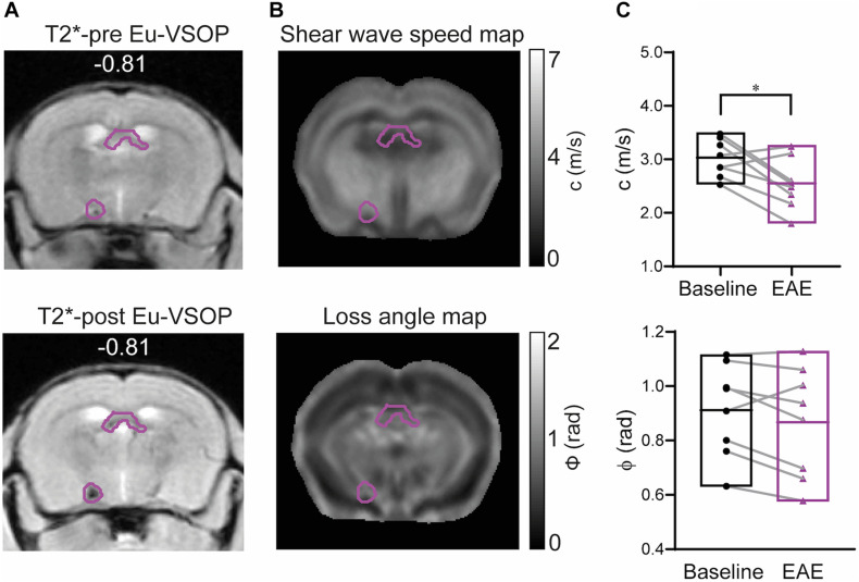
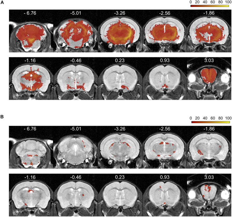
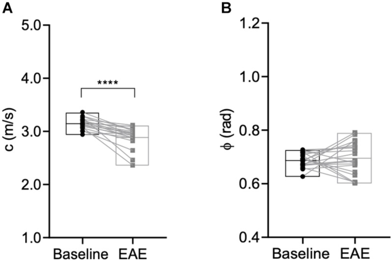
Neuroinflammatory processes occurring during multiple sclerosis cause disseminated softening of brain tissue, as quantified by magnetic resonance elastography (MRE). However, inflammation-mediated tissue alterations underlying the mechanical integrity of the brain remain unclear. We previously showed that blood-brain barrier (BBB) disruption visualized by MRI using gadolinium-based contrast agent (GBCA) does not correlate with tissue softening in active experimental autoimmune encephalomyelitis (EAE). However, it is unknown how confined BBB changes and other inflammatory processes may determine local elasticity changes. Therefore, we aim to elucidate which inflammatory hallmarks are determinant for local viscoelastic changes observed in EAE brains. Hence, novel multifrequency MRE was applied in combination with GBCA-based MRI or very small superparamagnetic iron oxide particles (VSOPs) in female SJL mice with induced adoptive transfer EAE ( = 21). VSOPs were doped with europium (Eu-VSOPs) to facilitate the analysis. Accumulation of Eu-VSOPs, which was previously demonstrated to be sensitive to immune cell infiltration and ECM remodeling, was also found to be independent of GBCA enhancement. Following registration to a reference brain atlas, viscoelastic properties of the whole brain and areas visualized by either Gd or VSOP were quantified. MRE revealed marked disseminated softening across the whole brain in mice with established EAE (baseline: 3.1 ± 0.1 m/s vs. EAE: 2.9 ± 0.2 m/s, < 0.0001). A similar degree of softening was observed in sites of GBCA enhancement i.e., mainly within cerebral cortex and brain stem (baseline: 3.3 ± 0.4 m/s vs. EAE: 3.0 ± 0.5 m/s, = 0.018). However, locations in which only Eu-VSOP accumulated, mainly in fiber tracts (baseline: 3.0 ± 0.4 m/s vs. EAE: 2.6 ± 0.5 m/s, = 0.023), softening was more pronounced when compared to non-hypointense areas (percent change of stiffness for Eu-VSOP accumulation: -16.81 ± 16.49% vs. for non-hypointense regions: -5.85 ± 3.81%, = 0.048). Our findings suggest that multifrequency MRE is sensitive to differentiate between local inflammatory processes with a strong immune cell infiltrate that lead to VSOP accumulation, from disseminated inflammation and BBB leakage visualized by GBCA. These pathological events visualized by Eu-VSOP MRI and MRE may include gliosis, macrophage infiltration, alterations of endothelial matrix components, and/or extracellular matrix remodeling. MRE may therefore represent a promising imaging tool for non-invasive clinical assessment of different pathological aspects of neuroinflammation.
多发性硬化症期间发生的神经炎症过程会导致脑组织的弥漫性软化,这可通过磁共振弹性成像(MRE)进行量化。然而,炎症介导的脑组织机械完整性改变的潜在机制仍不清楚。我们之前的研究表明,在活动性实验性自身免疫性脑脊髓炎(EAE)中,使用钆基造影剂(GBCA)通过MRI观察到的血脑屏障(BBB)破坏与组织软化无关。然而,局限性血脑屏障变化和其他炎症过程如何决定局部弹性变化尚不清楚。因此,我们旨在阐明哪些炎症特征是EAE脑局部粘弹性变化的决定因素。因此,将新型多频MRE与基于GBCA的MRI或超小超顺磁性氧化铁颗粒(VSOP)联合应用于诱导过继转移EAE的雌性SJL小鼠(n = 21)。VSOP用铕(Eu-VSOP)掺杂以促进分析。之前已证明对免疫细胞浸润和细胞外基质重塑敏感的Eu-VSOP积累也被发现与GBCA增强无关。在与参考脑图谱配准后,对全脑以及通过钆或VSOP可视化的区域的粘弹性特性进行了量化。MRE显示,在已建立EAE的小鼠中,全脑存在明显的弥漫性软化(基线:3.1±0.1 m/s vs. EAE:2.9±0.2 m/s,P < 0.0001)。在GBCA增强的部位,即主要在大脑皮层和脑干内,观察到了类似程度的软化(基线:3.3±0.4 m/s vs. EAE:3.0±0.5 m/s,P = 0.018)。然而,仅Eu-VSOP积累的部位,主要在纤维束中(基线:3.0±0.4 m/s vs. EAE:2.6±0.5 m/s,P = 0.023),与非低信号区域相比,软化更为明显(Eu-VSOP积累部位的硬度百分比变化:-16.81±16.49% vs. 非低信号区域:-