Golusda Laura, Kühl Anja A, Lehmann Malte, Dahlke Katja, Mueller Susanne, Boehm-Sturm Philipp, Saatz Jessica, Traub Heike, Schnorr Joerg, Freise Christian, Taupitz Matthias, Biskup Karina, Blanchard Véronique, Klein Oliver, Sack Ingolf, Siegmund Britta, Paclik Daniela
Medical Department, Division of Gastroenterology, Infectiology and Rheumatology, Campus Benjamin Franklin, Charité-Universitätsmedizin Berlin, Corporate Member of Freie Universität Berlin and Humboldt-Universität zu Berlin, Berlin, Germany.
iPATH.Berlin, Campus Benjamin Franklin, Charité-Universitätsmedizin Berlin, Corporate Member of Freie Universität Berlin and Humboldt-Universität zu Berlin, Berlin, Germany.
Front Physiol. 2022 Jul 12;13:862212. doi: 10.3389/fphys.2022.862212. eCollection 2022.
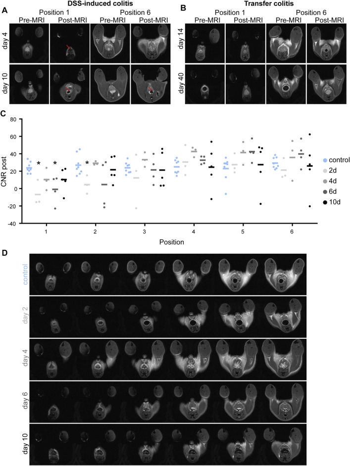
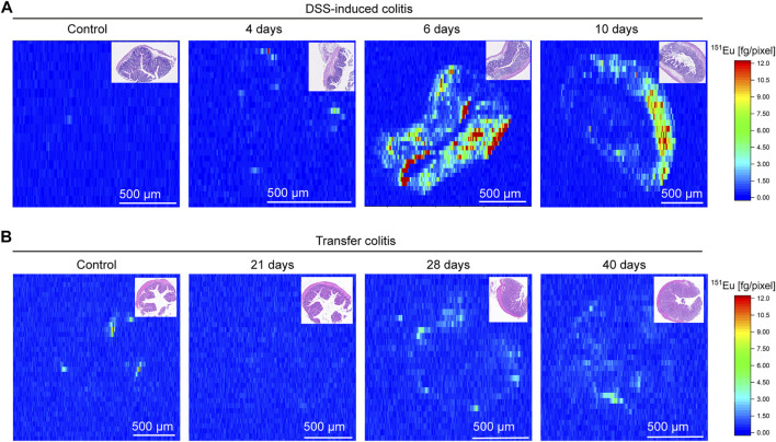
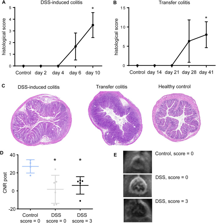
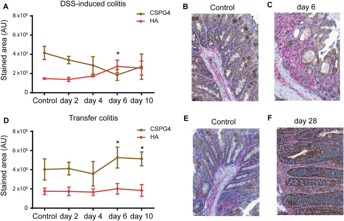
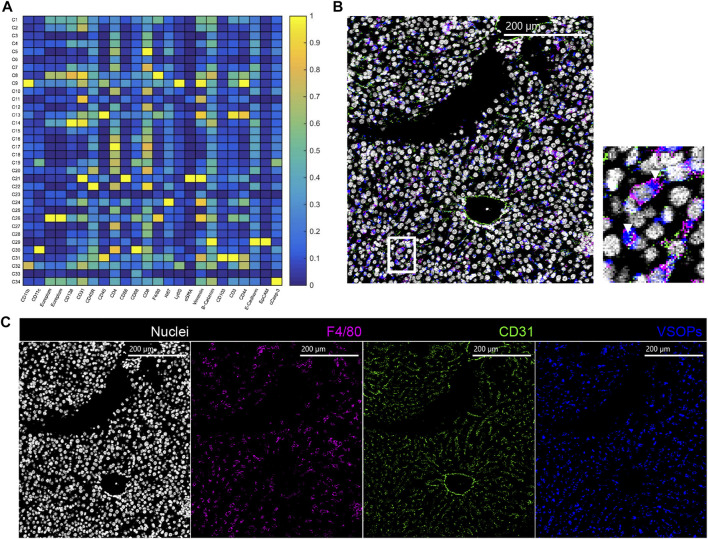
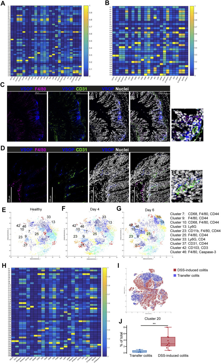
Inflammatory bowel diseases (IBD) comprise mainly ulcerative colitis (UC) and Crohn´s disease (CD). Both forms present with a chronic inflammation of the (gastro) intestinal tract, which induces excessive changes in the composition of the associated extracellular matrix (ECM). In UC, the inflammation is limited to the colon, whereas it can occur throughout the entire gastrointestinal tract in CD. Tools for early diagnosis of IBD are still very limited and highly invasive and measures for standardized evaluation of structural changes are scarce. To investigate an efficient non-invasive way of diagnosing intestinal inflammation and early changes of the ECM, very small superparamagnetic iron oxide nanoparticles (VSOPs) in magnetic resonance imaging (MRI) were applied in two mouse models of experimental colitis: the dextran sulfate sodium (DSS)-induced colitis and the transfer model of colitis. For further validation of ECM changes and inflammation, tissue sections were analyzed by immunohistochemistry. For in depth investigation of VSOPs localization within the tissue, Europium-doped VSOPs served to visualize the contrast agent by imaging mass cytometry (IMC). VSOPs accumulation in the inflamed colon wall of DSS-induced colitis mice was visualized in T* weighted MRI scans. Components of the ECM, especially the hyaluronic acid content, were found to influence VSOPs binding. Using IMC, co-localization of VSOPs with macrophages and endothelial cells in colon tissue was shown. In contrast to the DSS model, colonic inflammation could not be visualized with VSOP-enhanced MRI in transfer colitis. VSOPs present a potential contrast agent for contrast-enhanced MRI to detect intestinal inflammation in mice at an early stage and in a less invasive manner depending on hyaluronic acid content.
炎症性肠病(IBD)主要包括溃疡性结肠炎(UC)和克罗恩病(CD)。这两种形式均表现为(胃)肠道的慢性炎症,会导致相关细胞外基质(ECM)的组成发生过度变化。在UC中,炎症仅限于结肠,而在CD中,炎症可发生于整个胃肠道。IBD的早期诊断工具仍然非常有限且具有高度侵入性,用于标准化评估结构变化的措施也很少。为了研究一种诊断肠道炎症和ECM早期变化的有效非侵入性方法,在两种实验性结肠炎小鼠模型中应用了磁共振成像(MRI)中的超小超顺磁性氧化铁纳米颗粒(VSOP):葡聚糖硫酸钠(DSS)诱导的结肠炎和结肠炎转移模型。为了进一步验证ECM变化和炎症,通过免疫组织化学分析组织切片。为了深入研究VSOP在组织内的定位,掺铕VSOP用于通过成像质谱流式细胞术(IMC)可视化造影剂。在T*加权MRI扫描中可以看到VSOP在DSS诱导的结肠炎小鼠炎症结肠壁中的积聚。发现ECM的成分,尤其是透明质酸含量,会影响VSOP的结合。使用IMC显示了VSOP与结肠组织中的巨噬细胞和内皮细胞的共定位。与DSS模型不同,在转移型结肠炎中,VSOP增强的MRI无法显示结肠炎症。VSOP是一种潜在的造影剂,可用于增强MRI,以根据透明质酸含量以微创方式早期检测小鼠的肠道炎症。