Department of Radiation Oncology, Washington University School of Medicine, 4921 Parkview Place, Saint Louis MO 63110, USA.
BMC Med Imaging. 2013 Mar 2;13:7. doi: 10.1186/1471-2342-13-7.
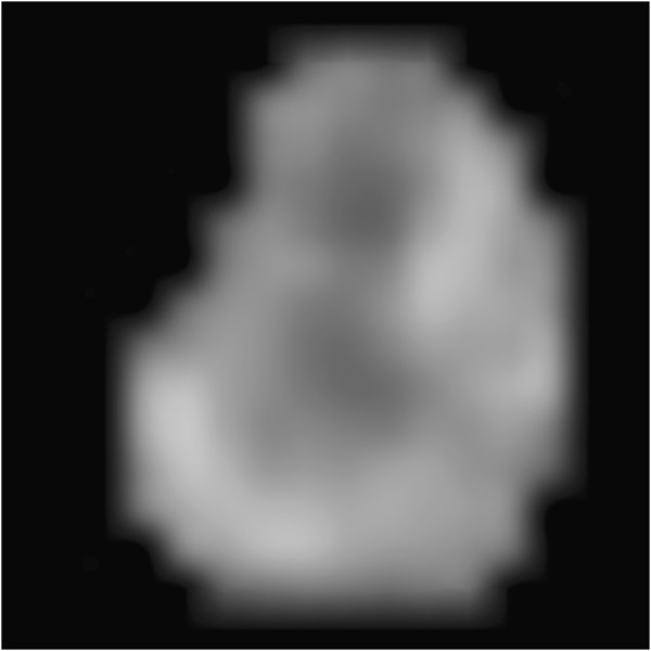
There has been much recent interest in the quantification of visually evident heterogeneity within functional grayscale medical images, such as those obtained via magnetic resonance or positron emission tomography. In the case of images of cancerous tumors, variations in grayscale intensity imply variations in crucial tumor biology. Despite these considerable clinical implications, there is as yet no standardized method for measuring the heterogeneity observed via these imaging modalities.
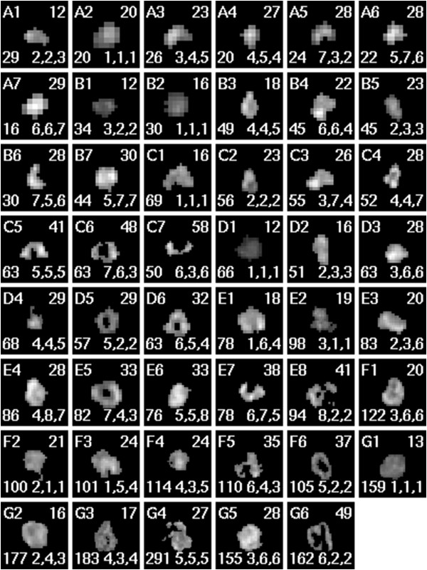
In this work, we motivate and derive a statistical measure of image heterogeneity. This statistic measures the distance-dependent average deviation from the smoothest intensity gradation feasible. We show how this statistic may be used to automatically rank images of in vivo human tumors in order of increasing heterogeneity. We test this method against the current practice of ranking images via expert visual inspection.
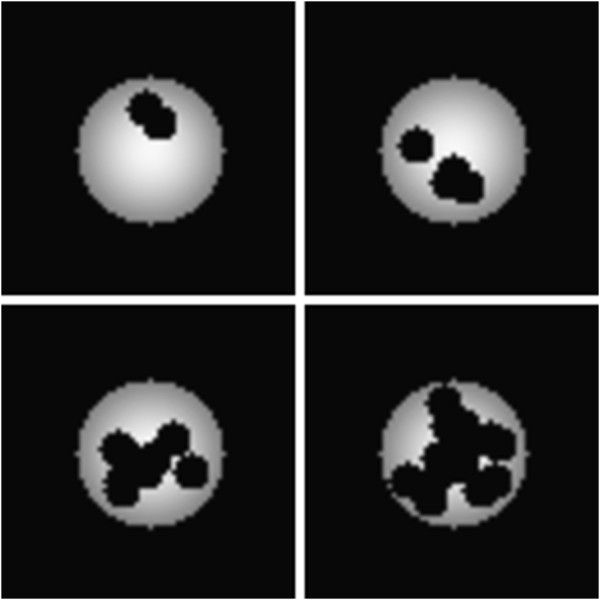
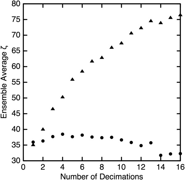
We find that this statistic provides a means of heterogeneity quantification beyond that given by other statistics traditionally used for the same purpose. We demonstrate the effect of tumor shape upon our ranking method and find the method applicable to a wide variety of clinically relevant tumor images. We find that the automated heterogeneity rankings agree very closely with those performed visually by experts.
These results indicate that our automated method may be used reliably to rank, in order of increasing heterogeneity, tumor images whether or not object shape is considered to contribute to that heterogeneity. Automated heterogeneity ranking yields objective results which are more consistent than visual rankings. Reducing variability in image interpretation will enable more researchers to better study potential clinical implications of observed tumor heterogeneity.
最近人们对功能灰度医学图像(如通过磁共振或正电子发射断层扫描获得的图像)中明显的异质性进行了大量的定量研究。在癌症肿瘤的图像中,灰度强度的变化意味着肿瘤生物学的关键变化。尽管这些都具有重要的临床意义,但目前还没有一种标准化的方法来测量这些成像方式观察到的异质性。
在这项工作中,我们提出并推导了一种用于测量图像异质性的统计量。该统计量衡量的是与最平滑的灰度渐变的距离相关的平均偏差。我们展示了如何使用该统计量自动对体内人类肿瘤图像进行排名,以表示异质性的增加。我们通过专家视觉检查来对这种方法和当前的图像排名方法进行比较。
我们发现,与传统用于相同目的的其他统计量相比,该统计量提供了一种异质性量化的方法。我们展示了肿瘤形状对我们的排名方法的影响,并发现该方法适用于广泛的临床相关肿瘤图像。我们发现,自动异质性排名与专家进行的视觉排名非常吻合。
这些结果表明,我们的自动方法可以可靠地用于对肿瘤图像进行排序,以便按照异质性的增加顺序进行排序,无论是否考虑物体形状对异质性的贡献。自动异质性排名产生的结果比视觉排名更一致。减少图像解释的可变性将使更多的研究人员能够更好地研究观察到的肿瘤异质性的潜在临床意义。