• 文献检索
  • 文档翻译
  • 深度研究
  • 学术资讯
  • Suppr Zotero 插件Zotero 插件
  • 邀请有礼
  • 套餐&价格
  • 历史记录
应用&插件
Suppr Zotero 插件Zotero 插件浏览器插件Mac 客户端Windows 客户端微信小程序
定价
高级版会员购买积分包购买API积分包
服务
文献检索文档翻译深度研究API 文档MCP 服务
关于我们
关于 Suppr公司介绍联系我们用户协议隐私条款
关注我们

Suppr 超能文献

核心技术专利:CN118964589B侵权必究
粤ICP备2023148730 号-1Suppr @ 2026

文献检索

告别复杂PubMed语法,用中文像聊天一样搜索,搜遍4000万医学文献。AI智能推荐,让科研检索更轻松。

立即免费搜索

文件翻译

保留排版,准确专业,支持PDF/Word/PPT等文件格式,支持 12+语言互译。

免费翻译文档

深度研究

AI帮你快速写综述,25分钟生成高质量综述,智能提取关键信息,辅助科研写作。

立即免费体验

磁共振成像显示颈动脉斑块出血强烈预示着复发性缺血和中风。

Carotid plaque hemorrhage on magnetic resonance imaging strongly predicts recurrent ischemia and stroke.

机构信息

Division of Radiological and Imaging Sciences, University of Nottingham, Queen's Medical Campus, Nottingham, United Kingdom.

出版信息

Ann Neurol. 2013 Jun;73(6):774-84. doi: 10.1002/ana.23876. Epub 2013 Jun 4.

DOI:10.1002/ana.23876
PMID:23463579
原文链接:https://pmc.ncbi.nlm.nih.gov/articles/PMC3824333/
Abstract

OBJECTIVE

There is a recognized need to improve selection of patients with carotid artery stenosis for carotid endarterectomy (CEA). We assessed the value of magnetic resonance imaging (MRI)-defined carotid plaque hemorrhage (MRIPH) to predict recurrent ipsilateral cerebral ischemic events, and stroke in symptomatic carotid stenosis.

METHODS

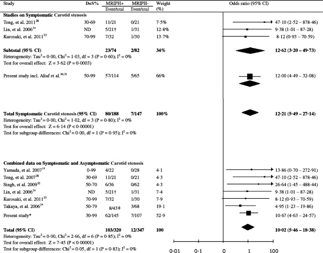
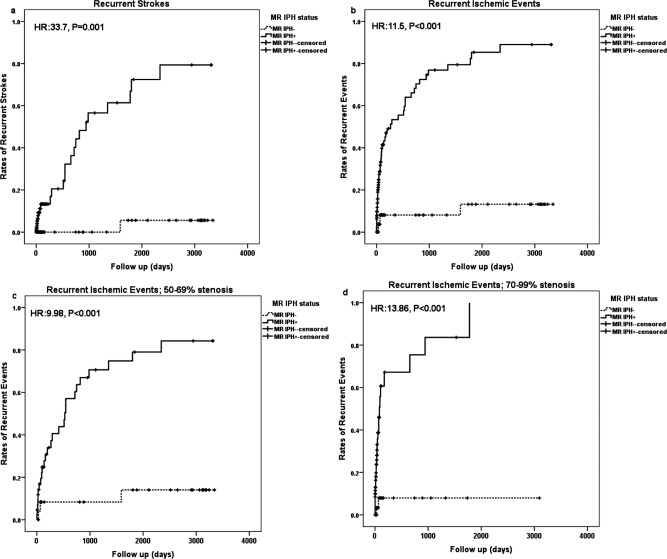
One hundred seventy-nine symptomatic patients with ≥ 50% stenosis were prospectively recruited, underwent carotid MRI, and were clinically followed up until CEA, death, or ischemic event. MRIPH was diagnosed if the plaque signal intensity was >150% that of the adjacent muscle. Event-free survival analysis was done using Kaplan-Meier plots and Cox regression models controlling for known vascular risk factors. We also undertook a meta-analysis of reported data on MRIPH and recurrent events.

RESULTS

One hundred fourteen patients (63.7%) showed MRIPH, suffering 92% (57 of 62) of all recurrent ipsilateral events and all but 1 (25 of 26) future strokes. Patients without MRIPH had an estimated annual absolute stroke risk of only 0.6%. Cox multivariate regression analysis proved MRIPH as a strong predictor of recurrent ischemic events (hazard ratio [HR] = 12.0, 95% confidence interval [CI] = 4.8-30.1, p < 0.001) and stroke alone (HR = 35.0, 95% CI = 4.7-261.6, p = 0.001). Meta-analysis of published data confirmed this association between MRIPH and recurrent cerebral ischemic events in symptomatic carotid artery stenosis (odds ratio = 12.2, 95% CI = 5.5-27.1, p < 0.00001).

INTERPRETATION

MRIPH independently and strongly predicts recurrent ipsilateral ischemic events, and stroke alone, in symptomatic ≥ 50% carotid artery stenosis. The very low stroke risk in patients without MRIPH puts into question current risk-benefit assessment for CEA in this subgroup.

摘要

目的

人们已经认识到需要改进颈动脉狭窄患者行颈动脉内膜切除术(CEA)的选择。我们评估了磁共振成像(MRI)定义的颈动脉斑块出血(MRIPH)对预测同侧复发性脑缺血事件和症状性颈动脉狭窄性卒中的价值。

方法

前瞻性招募了 179 例≥50%狭窄的症状性患者,行颈动脉 MRI 检查,并进行临床随访,直至 CEA、死亡或发生缺血事件。如果斑块信号强度比相邻肌肉高 150%,则诊断为 MRIPH。采用 Kaplan-Meier 图和 Cox 回归模型进行无事件生存分析,控制已知血管危险因素。我们还对 MRIPH 与复发性事件的报告数据进行了荟萃分析。

结果

114 例(63.7%)患者出现 MRIPH,发生了所有同侧复发性事件的 92%(57/62)和所有但 1 例(26/26)未来卒中。无 MRIPH 的患者估计每年的绝对卒中风险仅为 0.6%。Cox 多变量回归分析证明,MRIPH 是复发性缺血事件(危险比[HR] = 12.0,95%置信区间[CI] = 4.8-30.1,p < 0.001)和单独卒中的强烈预测因子(HR = 35.0,95% CI = 4.7-261.6,p = 0.001)。对已发表数据的荟萃分析证实了 MRIPH 与症状性颈动脉狭窄复发性脑缺血事件之间的这种关联(比值比= 12.2,95%CI = 5.5-27.1,p < 0.00001)。

结论

MRIPH 可独立且强烈预测症状性≥50%颈动脉狭窄患者的同侧复发性缺血事件和单独卒中。无 MRIPH 的患者卒中风险极低,这对该亚组患者行 CEA 的风险效益评估提出了质疑。

https://cdn.ncbi.nlm.nih.gov/pmc/blobs/9c42/3824333/e53b4e3b75b7/ana0073-0774-f3.jpg
https://cdn.ncbi.nlm.nih.gov/pmc/blobs/9c42/3824333/66b9e5f95d92/ana0073-0774-f1.jpg
https://cdn.ncbi.nlm.nih.gov/pmc/blobs/9c42/3824333/9716116cef5b/ana0073-0774-f2.jpg
https://cdn.ncbi.nlm.nih.gov/pmc/blobs/9c42/3824333/e53b4e3b75b7/ana0073-0774-f3.jpg
https://cdn.ncbi.nlm.nih.gov/pmc/blobs/9c42/3824333/66b9e5f95d92/ana0073-0774-f1.jpg
https://cdn.ncbi.nlm.nih.gov/pmc/blobs/9c42/3824333/9716116cef5b/ana0073-0774-f2.jpg
https://cdn.ncbi.nlm.nih.gov/pmc/blobs/9c42/3824333/e53b4e3b75b7/ana0073-0774-f3.jpg

相似文献

1
Carotid plaque hemorrhage on magnetic resonance imaging strongly predicts recurrent ischemia and stroke.磁共振成像显示颈动脉斑块出血强烈预示着复发性缺血和中风。
Ann Neurol. 2013 Jun;73(6):774-84. doi: 10.1002/ana.23876. Epub 2013 Jun 4.
2
Magnetic Resonance Imaging Plaque Hemorrhage for Risk Stratification in Carotid Artery Disease With Moderate Risk Under Current Medical Therapy.磁共振成像斑块出血用于当前药物治疗下中度风险颈动脉疾病的风险分层
Stroke. 2017 Mar;48(3):678-685. doi: 10.1161/STROKEAHA.116.015504. Epub 2017 Feb 14.
3
Extracranial carotid plaque hemorrhage predicts ipsilateral stroke recurrence in patients with carotid atherosclerosis - a study based on high-resolution vessel wall imaging MRI.基于高分辨率血管壁成像 MRI 的研究:颅外颈动脉斑块出血可预测颈动脉粥样硬化患者同侧卒中复发。
BMC Neurol. 2022 Jun 28;22(1):237. doi: 10.1186/s12883-022-02758-3.
4
Risk factors associated with cerebrovascular recurrence in symptomatic carotid disease: a comparative study of carotid plaque morphology, microemboli assessment and the European Carotid Surgery Trial risk model.有症状颈动脉疾病中与脑血管复发相关的危险因素:颈动脉斑块形态、微栓子评估及欧洲颈动脉外科试验风险模型的比较研究
J Am Heart Assoc. 2014 Jun 3;3(3):e000173. doi: 10.1161/JAHA.113.000173.
5
Moderate carotid artery stenosis: MR imaging-depicted intraplaque hemorrhage predicts risk of cerebrovascular ischemic events in asymptomatic men.中度颈动脉狭窄:磁共振成像显示的斑块内出血可预测无症状男性发生脑血管缺血事件的风险。
Radiology. 2009 Aug;252(2):502-8. doi: 10.1148/radiol.2522080792. Epub 2009 Jun 9.
6
Plaque At RISK (PARISK): prospective multicenter study to improve diagnosis of high-risk carotid plaques.易损斑块研究(PARISK):一项旨在改善高危颈动脉斑块诊断的前瞻性多中心研究。
Int J Stroke. 2014 Aug;9(6):747-54. doi: 10.1111/ijs.12167. Epub 2013 Oct 21.
7
Prediction of Stroke Risk by Detection of Hemorrhage in Carotid Plaques: Meta-Analysis of Individual Patient Data.基于颈动脉斑块出血检测预测卒中风险:个体患者数据分析的荟萃分析。
JACC Cardiovasc Imaging. 2020 Feb;13(2 Pt 1):395-406. doi: 10.1016/j.jcmg.2019.03.028. Epub 2019 Jun 12.
8
Signal intensity and volume of carotid intraplaque hemorrhage on magnetic resonance imaging and the risk of ipsilateral cerebrovascular events: The Plaque At RISK (PARISK) study.磁共振成像中颈动脉斑块内出血的信号强度和体积与同侧脑血管事件风险:斑块风险(PARISK)研究
J Cardiovasc Magn Reson. 2024;26(2):101049. doi: 10.1016/j.jocmr.2024.101049. Epub 2024 Jun 13.
9
Association of Progression of Carotid Artery Wall Volume and Recurrent Transient Ischemic Attack or Stroke: A Magnetic Resonance Imaging Study.颈动脉壁体积进展与复发性短暂性脑缺血发作或卒中的相关性:一项磁共振成像研究。
Stroke. 2018 Mar;49(3):614-620. doi: 10.1161/STROKEAHA.117.019422. Epub 2018 Jan 30.
10
Detection of intraplaque hemorrhage by magnetic resonance imaging in symptomatic patients with mild to moderate carotid stenosis predicts recurrent neurological events.在有轻至中度颈动脉狭窄的有症状患者中,通过磁共振成像检测斑块内出血可预测复发性神经事件。
J Vasc Surg. 2008 Feb;47(2):337-42. doi: 10.1016/j.jvs.2007.09.064.

引用本文的文献

1
Application of the Neuroform Atlas Stent for Intracranial Stenting of Symptomatic Intracranial Atherosclerotic Stenosis: A Multi-Center Retrospective Study.Neuroform Atlas支架在症状性颅内动脉粥样硬化狭窄颅内支架置入中的应用:一项多中心回顾性研究。
Brain Behav. 2025 Sep;15(9):e70798. doi: 10.1002/brb3.70798.
2
Risk of acute cerebral infarction in patients with hypertension based on high-resolution MRI.基于高分辨率磁共振成像的高血压患者急性脑梗死风险
Quant Imaging Med Surg. 2025 Apr 1;15(4):3000-3010. doi: 10.21037/qims-24-2106. Epub 2025 Mar 28.
3
An interpretable machine learning model for stroke recurrence in patients with symptomatic intracranial atherosclerotic arterial stenosis.

本文引用的文献

1
Magnetic resonance detected carotid plaque hemorrhage is associated with inflammatory features in symptomatic carotid plaques.磁共振检测到的颈动脉斑块内出血与有症状颈动脉斑块的炎症特征相关。
Ann Vasc Surg. 2013 Jul;27(5):655-61. doi: 10.1016/j.avsg.2012.10.011. Epub 2013 Mar 26.
2
Lower prevalence of carotid plaque hemorrhage in women, and its mediator effect on sex differences in recurrent cerebrovascular events.女性颈动脉斑块出血的患病率较低,其对复发性脑血管事件的性别差异具有中介作用。
PLoS One. 2012;7(10):e47319. doi: 10.1371/journal.pone.0047319. Epub 2012 Oct 26.
3
Symptomatic low-grade carotid stenosis with intraplaque hemorrhage and expansive arterial remodeling is associated with a high relapse rate refractory to medical treatment.
一种用于有症状颅内动脉粥样硬化性动脉狭窄患者卒中复发的可解释机器学习模型。
Front Neurosci. 2024 Jan 8;17:1323270. doi: 10.3389/fnins.2023.1323270. eCollection 2023.
4
Biliverdin Reductase B Is a Plasma Biomarker for Intraplaque Hemorrhage and a Predictor of Ischemic Stroke in Patients with Symptomatic Carotid Atherosclerosis.胆红素还原酶 B 是斑块内出血的血浆生物标志物,也是症状性颈动脉粥样硬化患者缺血性脑卒中的预测因子。
Biomolecules. 2023 May 24;13(6):882. doi: 10.3390/biom13060882.
5
Stroke risk prediction by color Doppler ultrasound of carotid artery-based deep learning using Inception V3 and VGG-16.基于Inception V3和VGG-16深度学习的颈动脉彩色多普勒超声中风风险预测
Front Neurol. 2023 Feb 14;14:1111906. doi: 10.3389/fneur.2023.1111906. eCollection 2023.
6
The role of non-stenosing carotid artery plaques in embolic stroke of undetermined source, is it a silent offender? A review of literature.非狭窄颈动脉斑块在不明来源栓塞性卒中中的作用,它是沉默的罪犯吗?文献复习。
Interv Neuroradiol. 2024 Oct;30(5):759-767. doi: 10.1177/15910199221143172. Epub 2022 Nov 30.
7
Sex Differences in Carotid Atherosclerosis: A Systematic Review and Meta-Analysis.颈动脉粥样硬化的性别差异:系统评价和荟萃分析。
Stroke. 2023 Feb;54(2):315-326. doi: 10.1161/STROKEAHA.122.041046. Epub 2022 Nov 29.
8
The role of cross-sectional imaging of the extracranial and intracranial vasculature in embolic stroke of undetermined source.颅外和颅内血管系统的横断面成像在不明来源栓塞性卒中中的作用。
Front Neurol. 2022 Aug 26;13:982896. doi: 10.3389/fneur.2022.982896. eCollection 2022.
9
Extracranial carotid plaque hemorrhage predicts ipsilateral stroke recurrence in patients with carotid atherosclerosis - a study based on high-resolution vessel wall imaging MRI.基于高分辨率血管壁成像 MRI 的研究:颅外颈动脉斑块出血可预测颈动脉粥样硬化患者同侧卒中复发。
BMC Neurol. 2022 Jun 28;22(1):237. doi: 10.1186/s12883-022-02758-3.
10
Radiomics Nomogram for Predicting Stroke Recurrence in Symptomatic Intracranial Atherosclerotic Stenosis.用于预测症状性颅内动脉粥样硬化狭窄患者卒中复发的影像组学列线图
Front Neurosci. 2022 Apr 12;16:851353. doi: 10.3389/fnins.2022.851353. eCollection 2022.
症状性低级别颈动脉狭窄伴斑块内出血和动脉扩张性重构与药物治疗难治性高复发率相关。
Neurosurgery. 2012 May;70(5):1143-50; discussion 1150-1. doi: 10.1227/NEU.0b013e31823fe50b.
4
Relationships between recent intraplaque hemorrhage and stroke risk factors in patients with carotid stenosis: the HIRISC study.颈动脉狭窄患者近期斑块内出血与卒中危险因素的关系:HIRISC 研究。
Arterioscler Thromb Vasc Biol. 2012 Feb;32(2):492-9. doi: 10.1161/ATVBAHA.111.239335. Epub 2011 Nov 10.
5
Late stage complicated atheroma in low-grade stenotic carotid disease: MR imaging depiction--prevalence and risk factors.低级别狭窄性颈动脉疾病中晚期复杂动脉粥样硬化:MR 成像表现——患病率和危险因素。
Radiology. 2011 Sep;260(3):841-7. doi: 10.1148/radiol.11101652. Epub 2011 Jul 6.
6
What is the current status of invasive treatment of extracranial carotid artery disease?颅外颈动脉疾病的介入治疗现状如何?
Stroke. 2011 Jul;42(7):2080-5. doi: 10.1161/STROKEAHA.110.597708. Epub 2011 Jun 9.
7
In vivo MRI-based 3D mechanical stress-strain profiles of carotid plaques with juxtaluminal plaque haemorrhage: an exploratory study for the mechanism of subsequent cerebrovascular events.载瘤腔外斑块出血的颈动脉斑块体内 MRI 三维机械应变成型:探讨后续脑血管事件发生机制的探索性研究。
Eur J Vasc Endovasc Surg. 2011 Oct;42(4):427-33. doi: 10.1016/j.ejvs.2011.05.009.
8
2011 ASA/ACCF/AHA/AANN/AANS/ACR/ASNR/CNS/SAIP/SCAI/SIR/SNIS/SVM/SVS guideline on the management of patients with extracranial carotid and vertebral artery disease: a report of the American College of Cardiology Foundation/American Heart Association Task Force on Practice Guidelines, and the American Stroke Association, American Association of Neuroscience Nurses, American Association of Neurological Surgeons, American College of Radiology, American Society of Neuroradiology, Congress of Neurological Surgeons, Society of Atherosclerosis Imaging and Prevention, Society for Cardiovascular Angiography and Interventions, Society of Interventional Radiology, Society of NeuroInterventional Surgery, Society for Vascular Medicine, and Society for Vascular Surgery.2011年美国麻醉医师协会/美国心脏病学会基金会/美国心脏协会/美国神经学会护士协会/美国神经外科医师协会/美国放射学会/美国神经放射学会/神经外科医师大会/动脉粥样硬化影像与预防学会/心血管造影和介入学会/介入放射学会/神经介入外科学会/血管医学学会和血管外科学会关于颅外颈动脉和椎动脉疾病患者管理的指南:美国心脏病学会基金会/美国心脏协会实践指南工作组、美国中风协会、美国神经科学护士协会、美国神经外科医师协会、美国放射学会、美国神经放射学会、神经外科医师大会、动脉粥样硬化影像与预防学会、心血管造影和介入学会、介入放射学会、神经介入外科学会、血管医学学会和血管外科学会的报告
J Am Coll Cardiol. 2011 Feb 22;57(8):e16-94. doi: 10.1016/j.jacc.2010.11.006. Epub 2011 Feb 1.
9
Discriminating carotid atherosclerotic lesion severity by luminal stenosis and plaque burden: a comparison utilizing high-resolution magnetic resonance imaging at 3.0 Tesla.利用 3.0T 高分辨率磁共振成像区分颈动脉粥样硬化病变严重程度:管腔狭窄和斑块负荷的比较。
Stroke. 2011 Feb;42(2):347-53. doi: 10.1161/STROKEAHA.110.597328. Epub 2010 Dec 23.
10
Plaque hemorrhage is a marker of thromboembolic activity in patients with symptomatic carotid disease.斑块出血是症状性颈动脉疾病患者血栓栓塞活性的标志物。
Radiology. 2011 Feb;258(2):538-45. doi: 10.1148/radiol.10100198. Epub 2010 Dec 16.