Shao Yilei, Tao Aizhu, Jiang Hong, Shen Meixiao, Zhong Jianguang, Lu Fan, Wang Jianhua
Bascom Palmer Eye Institute, University of Miami, Miami, FL, 33136, USA ; School of Ophthalmology and Optometry, Wenzhou Medical College, Wenzhou, Zhejiang, China ; These authors contributed equally to this work.
Biomed Opt Express. 2013 Mar 1;4(3):466-80. doi: 10.1364/BOE.4.000466. Epub 2013 Feb 21.
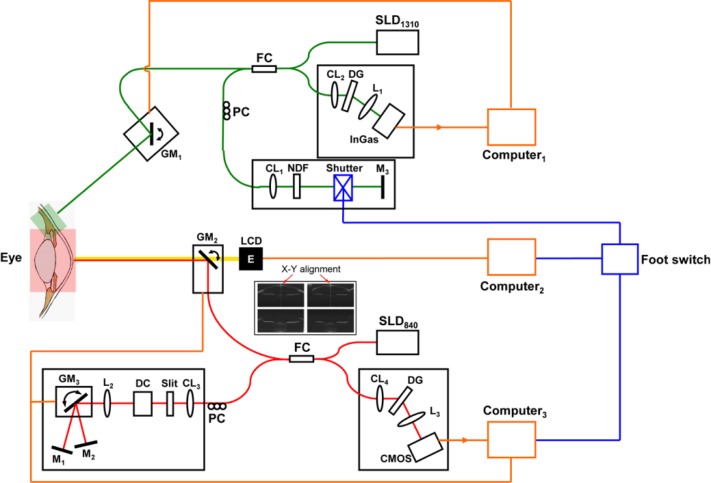
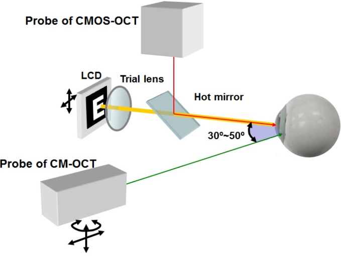
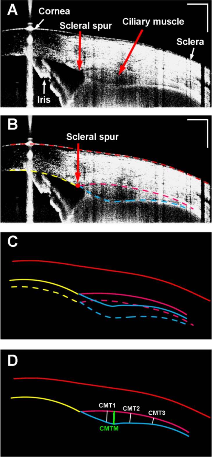
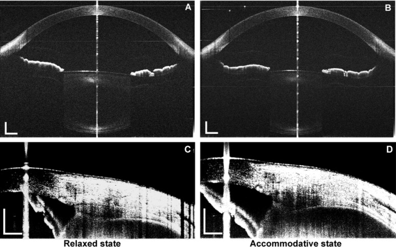
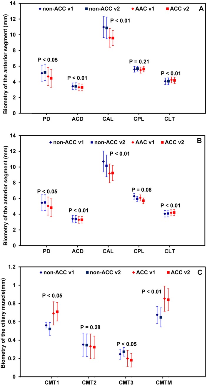
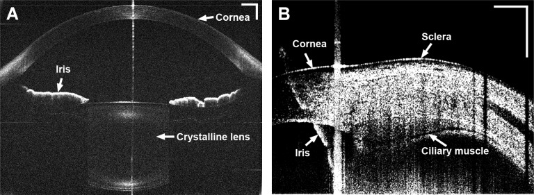
We demonstrated a novel approach of imaging the anterior segment including the ciliary muscle using combined and synchronized two spectral domain optical coherence tomography devices (SD-OCT). In one SD-OCT, a Complementary Metal-Oxide-Semiconductor Transistor (CMOS) camera and an alternating reference arm was used to image the anterior segment from the cornea to the lens. Another SD-OCT for imaging the ciliary muscle was equipped with a light source with a center wavelength of 1,310 nm and a bandwidth of 75 nm. Repeated measurements were performed under relaxed and 4.00 D accommodative stimulus states in six eyes from 6 subjects. We also imaged dynamic changes in the anterior segment in one eye during accommodation. The biometry of the anterior segment and the ciliary muscle was obtained. The combined system appeared to be capable to simultaneously real-time image the biometry of the anterior segment, including the ciliary muscle, in vivo during accommodation.
我们展示了一种使用组合式同步双光谱域光学相干断层扫描设备(SD-OCT)对包括睫状肌在内的眼前节进行成像的新方法。在一台SD-OCT中,使用互补金属氧化物半导体晶体管(CMOS)相机和交替参考臂对从角膜到晶状体的眼前节进行成像。另一台用于成像睫状肌的SD-OCT配备了中心波长为1310 nm、带宽为75 nm的光源。对6名受试者的6只眼睛在放松和4.00 D调节刺激状态下进行了重复测量。我们还对一只眼睛在调节过程中的眼前节动态变化进行了成像。获得了眼前节和睫状肌的生物测量数据。该组合系统似乎能够在调节过程中对包括睫状肌在内的眼前节生物测量数据进行体内实时同步成像。