Nanomedicine & Vision Group, Facultad de Ciencias Biomédicas, Universidad Austral, Pilar, Buenos Aires, Argentina.
BMC Ophthalmol. 2013 Apr 15;13:14. doi: 10.1186/1471-2415-13-14.
The contemporary peak of diabetes seems to be related to obesity, sedentary lifestyle and diet. Diabetic retinopathy is the most leading cause of blindness in adulthood in industrialized countries. Our purpose was to evaluate the effect of a high-fat-diet (HFD) on the retina of diabetic rats.
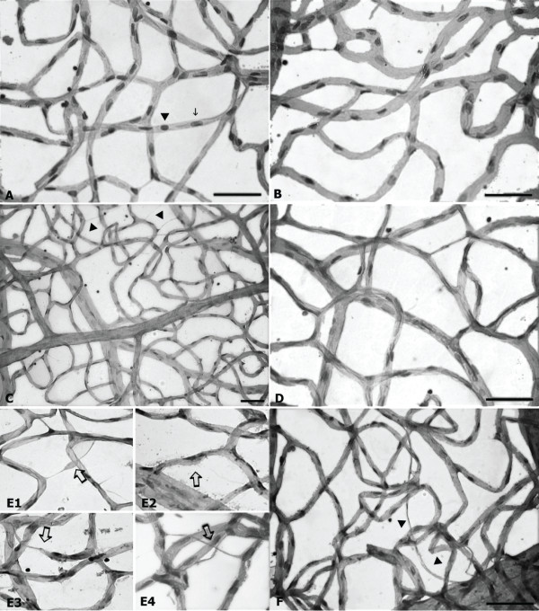
Two groups of Wistar rats were injected with streptozotocin (STZ) two days after birth using 45 and 90 mg/kg, respectively. At 8 weeks the group on lower doses started to be fed on a HFD. Animals were sacrificed at 37 weeks of diabetes. A control group was made up of non-diabetic rats. Retinal flat mounts were examined using the trypsin digestion technique. Pericytes counts were compared between diabetic and control rats. Cross retinal sections were analyzed by histological techniques and immunohistochemistry and immunofluorescent technique. Primary antibodies against inflammatory and proangiogenic mediators such as RAGE, GFAP, 5-LO, VEGF and TNF-α were used for immunohistochemistry and Western Blot (WB) analyses.
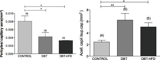
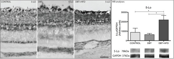
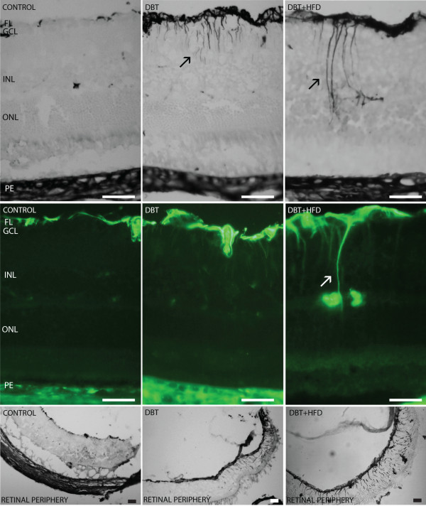
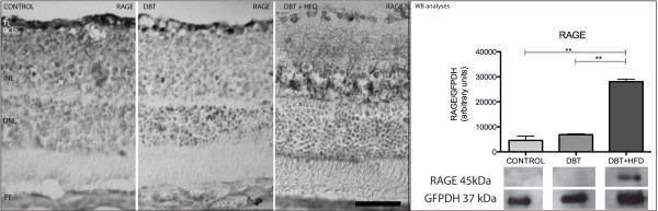
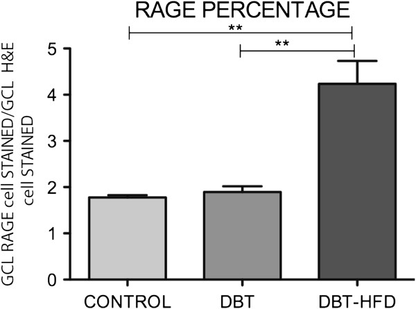
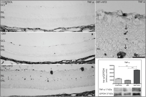
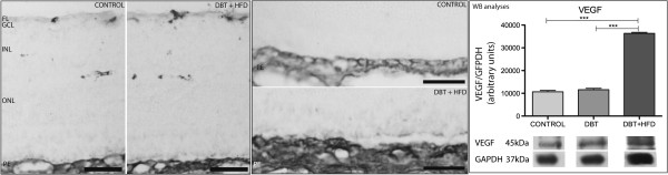
In the two diabetic groups we observed GFAP-positive cells with a morphology and spatial organization similar to those seen in Müller cells. Both diabetic groups had a significantly lower number of pericytes than non-diabetic animals.Increased retinal immunoreactivity of GFAP, RAGE, TNF-α, VEGF and 5-LO was seen in diabetic animals fed on HFD compared to the other groups of animals. WB analysis revealed a higher expression of 5-LO, VEGF, TNF-α and RAGE in the retina of diabetic rats on HFD than in controls and diabetics fed on a normal diet. The percentage of RAGE-stained ganglion cells and ganglion cells was found to be significantly lower in animals on a HFD than in the other animals.
Diabetic animals fed on a HFD showed an increased upregulation of inflammatory and proangiogenic markers. This animal model may be useful to study mechanisms of diabetic retinopathy and therapeutic targets.
当代糖尿病的高发似乎与肥胖、久坐不动的生活方式和饮食有关。糖尿病性视网膜病变是工业化国家成年人致盲的主要原因。我们的目的是评估高脂肪饮食(HFD)对糖尿病大鼠视网膜的影响。
两组 Wistar 大鼠在出生后两天分别用 45 和 90mg/kg 注射链脲佐菌素(STZ)。在较低剂量组的 8 周开始喂食 HFD。动物在糖尿病 37 周时被处死。对照组由非糖尿病大鼠组成。使用胰蛋白酶消化技术检查视网膜平片。比较糖尿病和对照组大鼠的周细胞计数。通过组织学技术和免疫组织化学及免疫荧光技术分析视网膜横切片。使用针对炎症和促血管生成介质(如 RAGE、GFAP、5-LO、VEGF 和 TNF-α)的一抗进行免疫组织化学和 Western blot(WB)分析。
在两个糖尿病组中,我们观察到 GFAP 阳性细胞具有与 Müller 细胞相似的形态和空间组织。与非糖尿病动物相比,两个糖尿病组的周细胞数量明显减少。与其他动物组相比,喂食 HFD 的糖尿病动物视网膜中 GFAP、RAGE、TNF-α、VEGF 和 5-LO 的免疫反应性显著增加。WB 分析显示,喂食 HFD 的糖尿病大鼠视网膜中 5-LO、VEGF、TNF-α和 RAGE 的表达明显高于对照组和正常饮食组的糖尿病大鼠。HFD 组的 RAGE 染色神经节细胞和神经节细胞的比例明显低于其他组。
喂食 HFD 的糖尿病动物表现出炎症和促血管生成标志物的上调增加。这种动物模型可能有助于研究糖尿病性视网膜病变的发病机制和治疗靶点。