Department of Materials, Imperial College London, London, UK.
Nat Mater. 2013 Jun;12(6):576-83. doi: 10.1038/nmat3627. Epub 2013 Apr 21.
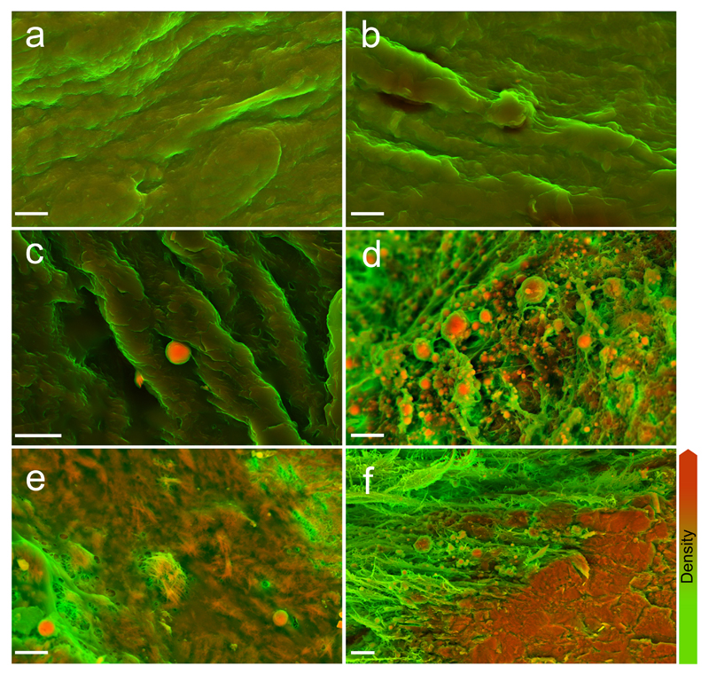
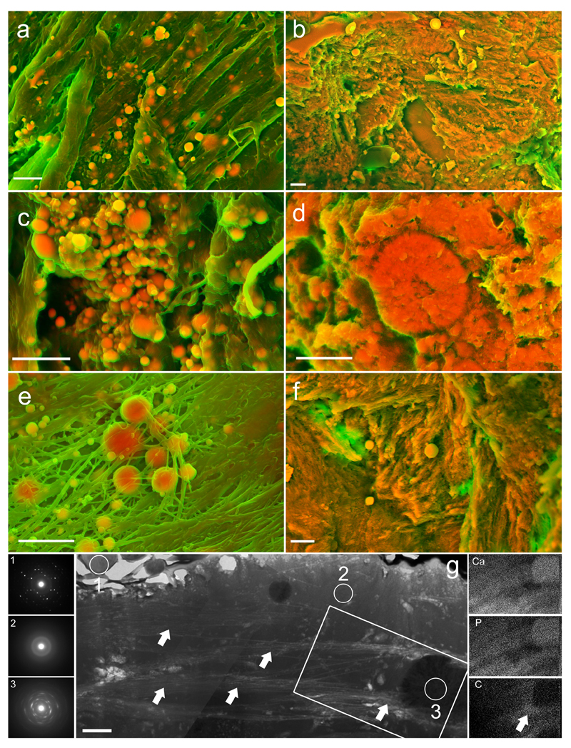
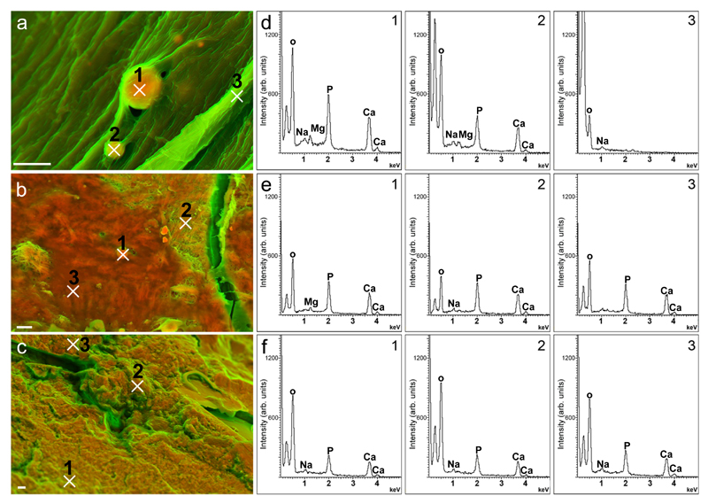
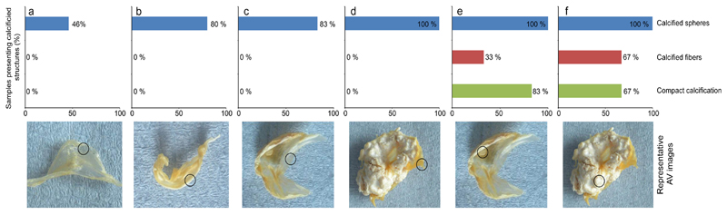
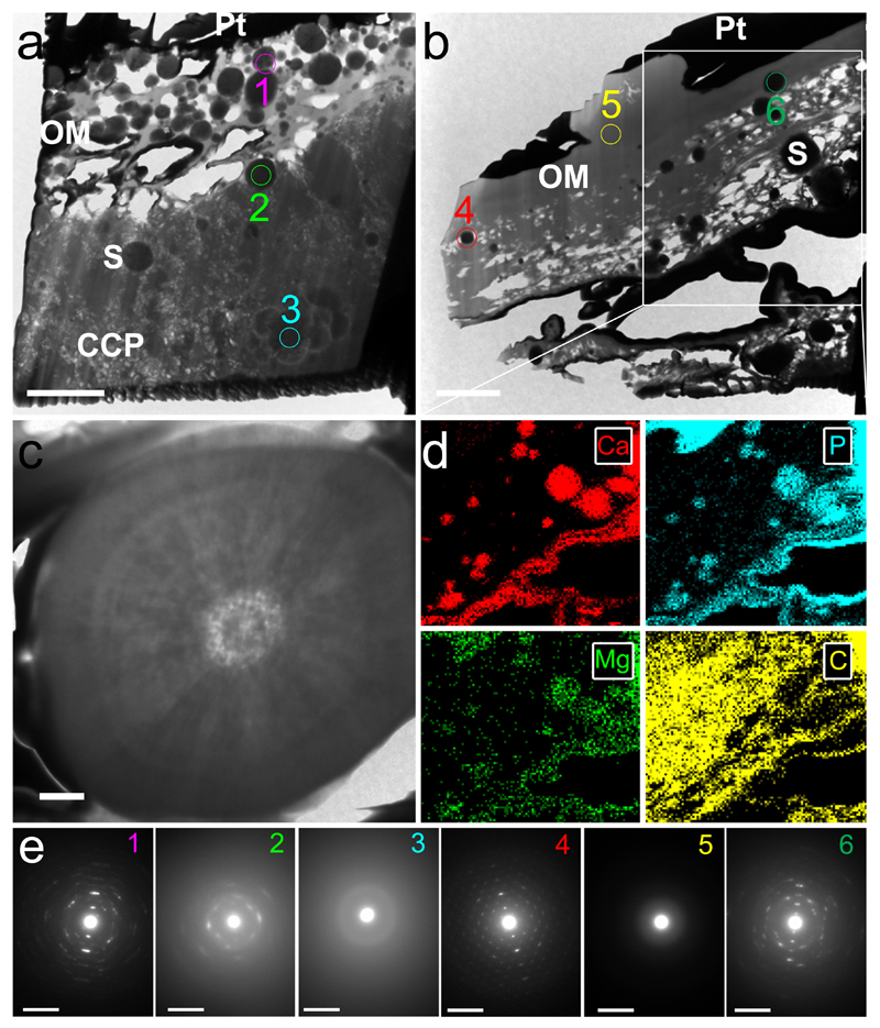
The accumulation of calcified material in cardiovascular tissue is thought to involve cytochemical, extracellular matrix and systemic signals; however, its precise composition and nanoscale architecture remain largely unexplored. Using nano-analytical electron microscopy techniques, we examined valves, aortae and coronary arteries from patients with and without calcific cardiovascular disease and detected spherical calcium phosphate particles, regardless of the presence of calcific lesions. We also examined lesions after sectioning with a focused ion beam and found that the spherical particles are composed of highly crystalline hydroxyapatite that crystallographically and structurally differs from bone mineral. Taken together, these data suggest that mineralized spherical particles may play a fundamental role in calcific lesion formation. Their ubiquitous presence in varied cardiovascular tissues and from patients with a spectrum of diseases further suggests that lesion formation may follow a common process. Indeed, applying materials science techniques to ectopic and orthotopic calcification has great potential to lend critical insights into pathophysiological processes underlying calcific cardiovascular disease.
心血管组织中钙化物质的积累被认为涉及细胞化学、细胞外基质和全身信号;然而,其确切的组成和纳米级结构在很大程度上仍未得到探索。使用纳米分析电子显微镜技术,我们检查了患有和不患有钙化性心血管疾病的患者的瓣膜、主动脉和冠状动脉,并检测到了球形磷酸钙颗粒,无论是否存在钙化病变。我们还使用聚焦离子束对切片进行了检查,发现这些球形颗粒由高度结晶的羟基磷灰石组成,其晶体结构和结构与骨矿物质不同。总的来说,这些数据表明,矿化的球形颗粒可能在钙化病变的形成中发挥着基本作用。它们在各种心血管组织中普遍存在,并且存在于患有各种疾病的患者中,这进一步表明病变的形成可能遵循一个共同的过程。事实上,将材料科学技术应用于异位和原位钙化具有很大的潜力,可以为钙化性心血管疾病的病理生理过程提供关键的见解。