• 文献检索
  • 文档翻译
  • 深度研究
  • 学术资讯
  • Suppr Zotero 插件Zotero 插件
  • 邀请有礼
  • 套餐&价格
  • 历史记录
应用&插件
Suppr Zotero 插件Zotero 插件浏览器插件Mac 客户端Windows 客户端微信小程序
定价
高级版会员购买积分包购买API积分包
服务
文献检索文档翻译深度研究API 文档MCP 服务
关于我们
关于 Suppr公司介绍联系我们用户协议隐私条款
关注我们

Suppr 超能文献

核心技术专利:CN118964589B侵权必究
粤ICP备2023148730 号-1Suppr @ 2026

文献检索

告别复杂PubMed语法,用中文像聊天一样搜索,搜遍4000万医学文献。AI智能推荐,让科研检索更轻松。

立即免费搜索

文件翻译

保留排版,准确专业,支持PDF/Word/PPT等文件格式,支持 12+语言互译。

免费翻译文档

深度研究

AI帮你快速写综述,25分钟生成高质量综述,智能提取关键信息,辅助科研写作。

立即免费体验

基于离子铣削切片电子显微镜的骨超微结构模型。

A model for the ultrastructure of bone based on electron microscopy of ion-milled sections.

机构信息

Department of Materials Science and Engineering, McMaster University, Hamilton, Ontario, Canada.

出版信息

PLoS One. 2012;7(1):e29258. doi: 10.1371/journal.pone.0029258. Epub 2012 Jan 17.

DOI:10.1371/journal.pone.0029258
PMID:22272230
原文链接:https://pmc.ncbi.nlm.nih.gov/articles/PMC3260135/
Abstract

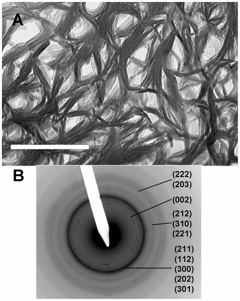
The relationship between the mineral component of bone and associated collagen has been a matter of continued dispute. We use transmission electron microscopy (TEM) of cryogenically ion milled sections of fully-mineralized cortical bone to study the spatial and topological relationship between mineral and collagen. We observe that hydroxyapatite (HA) occurs largely as elongated plate-like structures which are external to and oriented parallel to the collagen fibrils. Dark field images suggest that the structures ("mineral structures") are polycrystalline. They are approximately 5 nm thick, 70 nm wide and several hundred nm long. Using energy-dispersive X-ray analysis we show that approximately 70% of the HA occurs as mineral structures external to the fibrils. The remainder is found constrained to the gap zones. Comparative studies of other species suggest that this structural motif is ubiquitous in all vertebrates.

摘要

骨的矿物质成分与相关胶原之间的关系一直存在争议。我们使用低温离子铣切的完全矿化皮质骨切片的透射电子显微镜(TEM)来研究矿物质和胶原之间的空间和拓扑关系。我们观察到羟基磷灰石(HA)主要以长而薄的板状结构存在,这些结构位于胶原纤维之外,并与胶原纤维平行排列。暗场图像表明这些结构(“矿物质结构”)是多晶的。它们的厚度约为 5nm,宽度为 70nm,长度为数百纳米。通过能谱分析,我们发现大约 70%的 HA 以纤维外的矿物质结构存在。其余的则存在于纤维间隙中。对其他物种的比较研究表明,这种结构模式在所有脊椎动物中普遍存在。

https://cdn.ncbi.nlm.nih.gov/pmc/blobs/8aff/3260135/d34e10217338/pone.0029258.g010.jpg
https://cdn.ncbi.nlm.nih.gov/pmc/blobs/8aff/3260135/42aed1fadd7c/pone.0029258.g001.jpg
https://cdn.ncbi.nlm.nih.gov/pmc/blobs/8aff/3260135/d77118f2d670/pone.0029258.g002.jpg
https://cdn.ncbi.nlm.nih.gov/pmc/blobs/8aff/3260135/826233574591/pone.0029258.g003.jpg
https://cdn.ncbi.nlm.nih.gov/pmc/blobs/8aff/3260135/33c94321e022/pone.0029258.g004.jpg
https://cdn.ncbi.nlm.nih.gov/pmc/blobs/8aff/3260135/683aaa77f9f3/pone.0029258.g005.jpg
https://cdn.ncbi.nlm.nih.gov/pmc/blobs/8aff/3260135/ed9e0bf29374/pone.0029258.g006.jpg
https://cdn.ncbi.nlm.nih.gov/pmc/blobs/8aff/3260135/c56b873bf458/pone.0029258.g007.jpg
https://cdn.ncbi.nlm.nih.gov/pmc/blobs/8aff/3260135/64043716a911/pone.0029258.g008.jpg
https://cdn.ncbi.nlm.nih.gov/pmc/blobs/8aff/3260135/130fd39a7f4b/pone.0029258.g009.jpg
https://cdn.ncbi.nlm.nih.gov/pmc/blobs/8aff/3260135/d34e10217338/pone.0029258.g010.jpg

相似文献

1
A model for the ultrastructure of bone based on electron microscopy of ion-milled sections.基于离子铣削切片电子显微镜的骨超微结构模型。
PLoS One. 2012;7(1):e29258. doi: 10.1371/journal.pone.0029258. Epub 2012 Jan 17.
2
Dark-field transmission electron microscopy of cortical bone reveals details of extrafibrillar crystals.皮质骨的暗场透射电子显微镜检查揭示了纤维外晶体的细节。
J Struct Biol. 2014 Dec;188(3):240-8. doi: 10.1016/j.jsb.2014.10.005. Epub 2014 Oct 22.
3
Scanning transmission electron microscopic tomography of cortical bone using Z-contrast imaging.皮质骨的 Z 衬度成像扫描透射电子显微镜断层成像。
Micron. 2013 Jun;49:46-53. doi: 10.1016/j.micron.2013.03.002. Epub 2013 Mar 16.
4
The ultrastructure of bone as revealed in electron microscopy of ion-milled sections.离子研磨切片的电子显微镜观察所揭示的骨超微结构。
Semin Cell Dev Biol. 2015 Oct;46:44-50. doi: 10.1016/j.semcdb.2015.06.008. Epub 2015 Jul 9.
5
A search for apatite crystals in the gap zone of collagen fibrils in bone using dark-field illumination.使用暗场照明在骨胶原原纤维的间隙区寻找磷灰石晶体。
Bone. 2020 Jun;135:115304. doi: 10.1016/j.bone.2020.115304. Epub 2020 Mar 5.
6
Ultrastructure of Bone: Hierarchical Features from Nanometer to Micrometer Scale Revealed in Focused Ion Beam Sections in the TEM.骨的超微结构:在 TEM 中聚焦离子束切片中揭示的从纳米到微米尺度的层次特征。
Calcif Tissue Int. 2018 Dec;103(6):606-616. doi: 10.1007/s00223-018-0454-9. Epub 2018 Jul 14.
7
Average hydroxyapatite concentration is uniform in the extracollagenous ultrastructure of mineralized tissues: evidence at the 1-10-microm scale.矿化组织的胶原外超微结构中平均羟基磷灰石浓度是均匀的:1-10微米尺度的证据。
Biomech Model Mechanobiol. 2003 Aug;2(1):21-36. doi: 10.1007/s10237-002-0025-9.
8
Minerals and aligned collagen fibrils in tilapia fish scales: structural analysis using dark-field and energy-filtered transmission electron microscopy and electron tomography.罗非鱼鳞片中的矿物质和取向胶原纤维:暗场和能谱过滤透射电子显微镜及电子断层扫描的结构分析。
Microsc Microanal. 2011 Oct;17(5):788-98. doi: 10.1017/S1431927611011949. Epub 2011 Sep 8.
9
Identification of collagen fibrils in cross sections of bone by electron energy loss spectroscopy (EELS).通过电子能量损失谱(EELS)鉴定骨横截面上的胶原纤维。
Micron. 2019 Sep;124:102706. doi: 10.1016/j.micron.2019.102706. Epub 2019 Jun 20.
10
The locus of mineral crystallites in bone.骨中矿物质微晶的位点。
Connect Tissue Res. 1988;18(1):41-54. doi: 10.3109/03008208809019071.

引用本文的文献

1
Mineral and cross-linking in collagen fibrils: The mechanical behavior of bone tissue at the nano-scale.胶原原纤维中的矿物质和交联:纳米尺度下骨组织的力学行为。
J Mech Behav Biomed Mater. 2024 Nov;159:106697. doi: 10.1016/j.jmbbm.2024.106697. Epub 2024 Aug 22.
2
Bone hierarchical organization through the lens of materials science: Present opportunities and future challenges.透过材料科学视角看骨的层级组织:当前机遇与未来挑战
Bone Rep. 2024 Jun 25;22:101783. doi: 10.1016/j.bonr.2024.101783. eCollection 2024 Sep.
3
Elucidating the role of diverse mineralisation paradigms on bone biomechanics - a coarse-grained molecular dynamics investigation.

本文引用的文献

1
Deposition of apatite in mineralizing vertebrate extracellular matrices: A model of possible nucleation sites on type I collagen.矿化脊椎动物细胞外基质中磷灰石的沉积:I 型胶原上可能的成核位点模型。
Connect Tissue Res. 2011 Jun;52(3):242-54. doi: 10.3109/03008207.2010.551567. Epub 2011 Mar 15.
2
Hierarchical and non-hierarchical mineralisation of collagen.胶原的分级和非分级矿化。
Biomaterials. 2011 Feb;32(5):1291-300. doi: 10.1016/j.biomaterials.2010.10.018. Epub 2010 Oct 30.
3
The role of collagen in bone apatite formation in the presence of hydroxyapatite nucleation inhibitors.
阐明不同矿化模式对骨骼生物力学的作用——粗粒分子动力学研究。
Nanoscale. 2024 Feb 8;16(6):3173-3184. doi: 10.1039/d3nr04660e.
4
Ultrastructure and Nanoporosity of Human Bone Shown with Correlative On-Axis Electron and Spectroscopic Tomographies.人骨的超微结构和纳米多孔性通过共轴电子和光谱断层扫描显示。
ACS Nano. 2023 Dec 26;17(24):24710-24724. doi: 10.1021/acsnano.3c04633. Epub 2023 Oct 17.
5
A coarse-grained molecular dynamics investigation of the role of mineral arrangement on the mechanical properties of mineralized collagen fibrils.粗粒化分子动力学研究矿物质排列对矿化胶原原纤维力学性能的作用。
J R Soc Interface. 2023 Jan;20(198):20220803. doi: 10.1098/rsif.2022.0803. Epub 2023 Jan 25.
6
Advances in biomimetic collagen mineralisation and future approaches to bone tissue engineering.仿生胶原矿化研究进展及其在骨组织工程中的未来应用。
Biopolymers. 2023 Jan;114(1):e23527. doi: 10.1002/bip.23527. Epub 2022 Nov 29.
7
Hierarchical Structure and Properties of the Bone at Nano Level.纳米级骨的层次结构与特性
Bioengineering (Basel). 2022 Nov 10;9(11):677. doi: 10.3390/bioengineering9110677.
8
Perspective on Dentoalveolar Manifestations Resulting From PHOSPHO1 Loss-of-Function: A Form of Pseudohypophosphatasia?PHOSPHO1功能丧失导致的牙槽骨表现透视:一种假性低磷酸酯酶症形式?
Front Dent Med. 2022 Feb;3. doi: 10.3389/fdmed.2022.826387. Epub 2022 Feb 3.
9
Designed nanostructures created physicochemical switching of the growth mode between single crystals and mesocrystals.设计的纳米结构实现了单晶和中晶之间生长模式的物理化学切换。
Nanoscale Adv. 2022 Feb 18;4(6):1538-1544. doi: 10.1039/d1na00784j. eCollection 2022 Mar 15.
10
Preparation of hybrid samples for scanning electron microscopy (SEM) coupled to focused ion beam (FIB) analysis: A new way to study cell adhesion to titanium implant surfaces.用于扫描电子显微镜(SEM)与聚焦离子束(FIB)分析联用的混合样品制备:一种研究细胞与钛植入物表面黏附的新方法。
PLoS One. 2022 Aug 2;17(8):e0272486. doi: 10.1371/journal.pone.0272486. eCollection 2022.
胶原在存在羟磷灰石成核抑制剂时对骨磷灰石形成的作用。
Nat Mater. 2010 Dec;9(12):1004-9. doi: 10.1038/nmat2875. Epub 2010 Oct 24.
4
Biominerals--hierarchical nanocomposites: the example of bone.生物矿物--分级纳米复合材料:以骨骼为例。
Wiley Interdiscip Rev Nanomed Nanobiotechnol. 2011 Jan-Feb;3(1):47-69. doi: 10.1002/wnan.105.
5
The nano-morphological relationships between apatite crystals and collagen fibrils in ivory dentine.象牙质牙本质中磷灰石晶体与胶原原纤维的纳米形态关系。
Biomaterials. 2010 Jul;31(19):5275-86. doi: 10.1016/j.biomaterials.2010.03.025. Epub 2010 Apr 9.
6
Focused ion beam milling and ultramicrotomy of mineralised ivory dentine for analytical transmission electron microscopy.用于分析透射电子显微镜的矿化象牙质牙本质的聚焦离子束铣削和超薄切片术。
Micron. 2009 Jun;40(4):495-501. doi: 10.1016/j.micron.2008.12.002. Epub 2008 Dec 24.
7
Cooperative deformation of mineral and collagen in bone at the nanoscale.纳米尺度下骨中矿物质与胶原蛋白的协同变形
Proc Natl Acad Sci U S A. 2006 Nov 21;103(47):17741-6. doi: 10.1073/pnas.0604237103. Epub 2006 Nov 9.
8
Ultrastructural examination of dentin using focused ion-beam cross-sectioning and transmission electron microscopy.使用聚焦离子束横截面技术和透射电子显微镜对牙本质进行超微结构检查。
Micron. 2005;36(7-8):672-80. doi: 10.1016/j.micron.2005.05.011. Epub 2005 Sep 6.
9
Atomic force microscopic studies on the structure of bovine femoral cortical bone at the collagen fibril-mineral level.在胶原纤维 - 矿物质水平上对牛股骨皮质骨结构的原子力显微镜研究。
J Mater Sci Mater Med. 2002 Mar;13(3):333-7. doi: 10.1023/a:1014079421895.
10
Collagen-crystal relationships in bone as seen in the electron microscope.骨中胶原-晶体关系的电子显微镜观察
Anat Rec. 1952 Nov;114(3):383-409. doi: 10.1002/ar.1091140302.