Department of Neurology and Psychiatry, Sapienza University, Rome, Italy.
PLoS One. 2013 May 16;8(5):e63250. doi: 10.1371/journal.pone.0063250. Print 2013.
Multiple sclerosis (MS) is characterized by demyelinating and degenerative processes within the central nervous system. Unlike conventional MRI,new advanced imaging techniques improve pathological specificity and better highlight the relationship between anatomical damage and clinical impairment.
To investigate the relationship between clinical disability and both grey (GM) and white matter (WM) regional damage in MS patients.
Thirty-six relapsing remitting-MS patients and 25 sex- and age-matched controls were enrolled. All patients were clinically evaluated by the Expanded Disability Status Scale and the Multiple Sclerosis Functional Composite (MSFC) scale, which includes the 9-hole peg test (9HPT), the timed 25-feet walking test (T25FW) and the paced auditory serial addition test (PASAT). All subjects were imaged by a 3.0 T scanner: dual-echo fast spin-echo, 3DT1-weighted and diffusion-tensor imaging (DTI) sequences were acquired. Voxel-based morphometry (VBM) and tract-based spatial statistics (TBSS) analyses were run for regional GM and WM assessment, respectively. T2 lesion volumes were also calculated, by using a semi-automated technique.
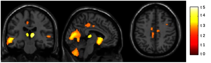
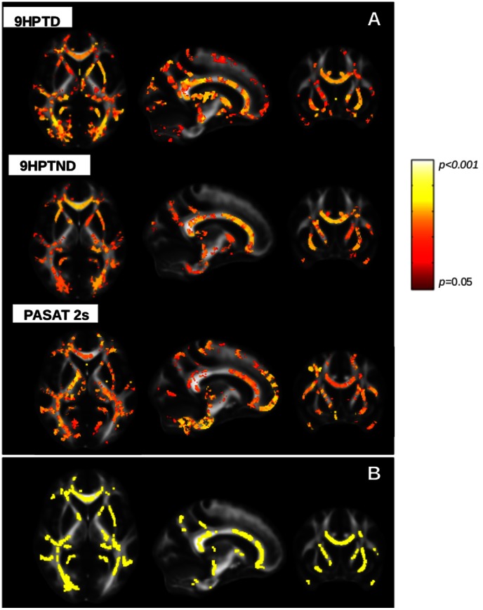
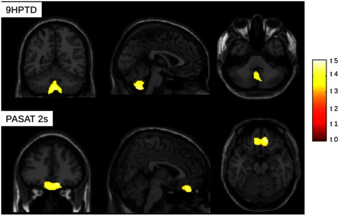
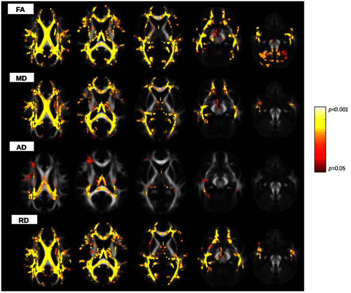
Brain volumetric assessment of GM and DTI measures revealed significant differences between patients and controls. In patients, different measures of WM damage correlated each-other (p<0.0001), whereas none of them correlated with GM volume. In patients, focal GM atrophy and widespread WM damage significantly correlated with clinical measures. In particular, VBM analysis revealed a significant correlation (p<0.05) between GM volume and 9HPT in cerebellum and between GM volume and PASAT in orbito-frontal cortex. TBSS showed significant correlations between DTI metrics with 9HPT and PASAT scores in many WM bundles (p<0.05), including corpus callosum, internal capsule, posterior thalamic radiations, cerebral peduncles.
Selective GM atrophy and widespread WM tracts damage are associated with functional impairment of upper-limb motion and cognition. The combined analysis of volumetric and DTI data may help to better understand structural alterations underlying physical and cognitive dysfunction in MS.
多发性硬化症(MS)的特征是中枢神经系统内的脱髓鞘和退行性过程。与传统 MRI 不同,新的高级成像技术提高了病理学特异性,并更好地突出了解剖损伤与临床损伤之间的关系。
探讨 MS 患者临床残疾与灰质(GM)和白质(WM)区域损伤之间的关系。
纳入 36 例复发缓解型 MS 患者和 25 名性别和年龄匹配的对照者。所有患者均通过扩展残疾状况量表和多发性硬化功能综合量表(MSFC)进行临床评估,其中包括 9 孔钉测试(9HPT)、定时 25 英尺步行测试(T25FW)和定速听觉序列加法测试(PASAT)。所有受试者均采用 3.0T 扫描仪进行成像:采集双回波快速自旋回波、3DT1 加权和弥散张量成像(DTI)序列。分别进行基于体素的形态计量学(VBM)和基于束的空间统计学(TBSS)分析,以评估 GM 和 WM 的区域变化。使用半自动技术计算 T2 病变体积。
GM 和 DTI 测量的脑容积评估显示患者和对照组之间存在显著差异。在患者中,不同的 WM 损伤测量值相互关联(p<0.0001),而与 GM 体积均无关联。在患者中,局灶性 GM 萎缩和广泛的 WM 损伤与临床指标显著相关。特别是,VBM 分析显示 GM 体积与小脑 9HPT 之间(p<0.05)以及眶额皮质与 PASAT 之间(p<0.05)存在显著相关性。TBSS 显示 DTI 指标与 9HPT 和 PASAT 评分在许多 WM 束(p<0.05)之间存在显著相关性,包括胼胝体、内囊、丘脑后辐射、大脑脚。
选择性 GM 萎缩和广泛的 WM 束损伤与上肢运动和认知功能的功能障碍有关。容积和 DTI 数据的联合分析可能有助于更好地理解 MS 中物理和认知功能障碍的结构改变。