• 文献检索
  • 文档翻译
  • 深度研究
  • 学术资讯
  • Suppr Zotero 插件Zotero 插件
  • 邀请有礼
  • 套餐&价格
  • 历史记录
应用&插件
Suppr Zotero 插件Zotero 插件浏览器插件Mac 客户端Windows 客户端微信小程序
定价
高级版会员购买积分包购买API积分包
服务
文献检索文档翻译深度研究API 文档MCP 服务
关于我们
关于 Suppr公司介绍联系我们用户协议隐私条款
关注我们

Suppr 超能文献

核心技术专利:CN118964589B侵权必究
粤ICP备2023148730 号-1Suppr @ 2026

文献检索

告别复杂PubMed语法,用中文像聊天一样搜索,搜遍4000万医学文献。AI智能推荐,让科研检索更轻松。

立即免费搜索

文件翻译

保留排版,准确专业,支持PDF/Word/PPT等文件格式,支持 12+语言互译。

免费翻译文档

深度研究

AI帮你快速写综述,25分钟生成高质量综述,智能提取关键信息,辅助科研写作。

立即免费体验

静息 CT 心肌灌注在疑似急性冠状动脉综合征患者中的诊断性能。

Diagnostic performance of resting CT myocardial perfusion in patients with possible acute coronary syndrome.

机构信息

Division of Cardiology, University of Washington, Seattle, WA 98195, USA.

出版信息

AJR Am J Roentgenol. 2013 May;200(5):W450-7. doi: 10.2214/AJR.12.8934.

DOI:10.2214/AJR.12.8934
PMID:23617513
原文链接:https://pmc.ncbi.nlm.nih.gov/articles/PMC4039350/
Abstract

OBJECTIVE

Coronary CT angiography has high sensitivity, but modest specificity, to detect acute coronary syndrome. We studied whether adding resting CT myocardial perfusion imaging improved the detection of acute coronary syndrome.

SUBJECTS AND METHODS

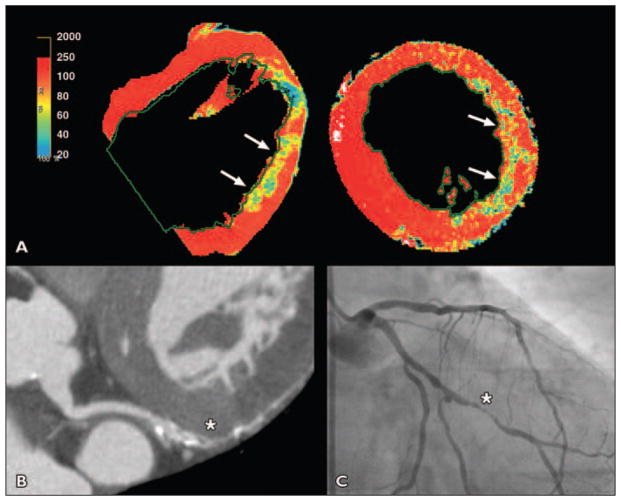
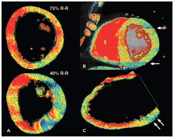
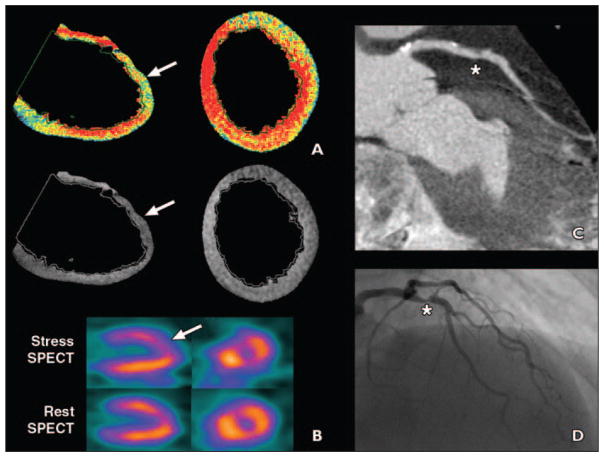
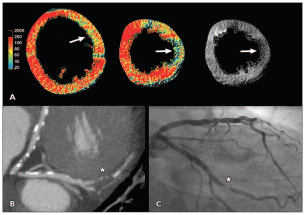
Patients with low-to-intermediate cardiac risk presenting with possible acute coronary syndrome received both the standard of care evaluation and a research thoracic 64-MDCT examination. Patients with an obstructive (> 50%) stenosis or a nonevaluable coronary segment on CT were diagnosed with possible acute coronary syndrome. CT perfusion was determined by applying gray and color Hounsfield unit maps to resting CT angiography images. Adjudicated patient diagnoses were based on the standard of care and 3-month follow-up. Patient-level diagnostic performance for acute coronary syndrome was calculated for coronary CT, CT perfusion, and combined techniques.

RESULTS

A total of 105 patients were enrolled. Of the nine (9%) patients with acute coronary syndrome, all had obstructive CT stenoses but only three had abnormal CT perfusion. CT perfusion was normal in all other patients. To detect acute coronary syndrome, CT angiography had 100% sensitivity, 89% specificity, and a positive predictive value of 45%. For CT perfusion, specificity and positive predictive value were each 100%, and sensitivity was 33%. Combined cardiac CT and CT perfusion had similar specificity but a higher positive predictive value (100%) than did CT angiography.

CONCLUSION

Resting CT perfusion using CT angiographic images may have high specificity and may improve CT positive predictive value for acute coronary syndrome without added radiation and contrast. However, normal resting CT perfusion cannot exclude acute coronary syndrome.

摘要

目的

冠状动脉 CT 血管造影术对检测急性冠状动脉综合征具有较高的敏感性,但特异性较低。我们研究了静息 CT 心肌灌注成像是否能提高急性冠状动脉综合征的检出率。

方法

低至中度心脏风险的患者出现可能的急性冠状动脉综合征时,接受标准护理评估和研究性胸部 64-MDCT 检查。CT 上有>50%的阻塞性狭窄或无法评估的冠状动脉节段的患者被诊断为可能的急性冠状动脉综合征。通过将灰度和彩色亨氏单位图应用于静息 CT 血管造影图像来确定 CT 灌注。经裁决的患者诊断结果基于标准护理和 3 个月的随访。计算冠状动脉 CT、CT 灌注和联合技术对急性冠状动脉综合征的患者水平诊断性能。

结果

共纳入 105 例患者。在 9%(9 例)的急性冠状动脉综合征患者中,所有患者均有阻塞性 CT 狭窄,但只有 3 例有异常 CT 灌注。所有其他患者的 CT 灌注均正常。为了检测急性冠状动脉综合征,CT 血管造影术的敏感性为 100%,特异性为 89%,阳性预测值为 45%。对于 CT 灌注,特异性和阳性预测值均为 100%,而敏感性为 33%。与 CT 血管造影术相比,联合心脏 CT 和 CT 灌注的特异性相似,但阳性预测值更高(100%)。

结论

使用 CT 血管造影图像的静息 CT 灌注可能具有较高的特异性,并可能提高 CT 对急性冠状动脉综合征的阳性预测值,而不会增加辐射和对比剂的使用。然而,正常的静息 CT 灌注并不能排除急性冠状动脉综合征。

https://cdn.ncbi.nlm.nih.gov/pmc/blobs/fc02/4039350/b6fccfaec1bd/nihms500137f5.jpg
https://cdn.ncbi.nlm.nih.gov/pmc/blobs/fc02/4039350/d46cca43a245/nihms500137f1.jpg
https://cdn.ncbi.nlm.nih.gov/pmc/blobs/fc02/4039350/47098565fbf9/nihms500137f2.jpg
https://cdn.ncbi.nlm.nih.gov/pmc/blobs/fc02/4039350/cd8c3c2a2c37/nihms500137f3.jpg
https://cdn.ncbi.nlm.nih.gov/pmc/blobs/fc02/4039350/ba5f80c8f2df/nihms500137f4.jpg
https://cdn.ncbi.nlm.nih.gov/pmc/blobs/fc02/4039350/b6fccfaec1bd/nihms500137f5.jpg

相似文献

1
Diagnostic performance of resting CT myocardial perfusion in patients with possible acute coronary syndrome.静息 CT 心肌灌注在疑似急性冠状动脉综合征患者中的诊断性能。
AJR Am J Roentgenol. 2013 May;200(5):W450-7. doi: 10.2214/AJR.12.8934.
2
Myocardial hypo-enhancement on resting computed tomography angiography images accurately identifies myocardial hypoperfusion.静息 CT 血管造影图像上的心肌低增强可准确识别心肌低灌注。
J Cardiovasc Comput Tomogr. 2011 Nov-Dec;5(6):412-20. doi: 10.1016/j.jcct.2011.10.006. Epub 2011 Oct 24.
3
Incremental value of myocardial perfusion over regional left ventricular function and coronary stenosis by cardiac CT for the detection of acute coronary syndromes in high-risk patients: a subgroup analysis of the ROMICAT trial.心脏 CT 心肌灌注与局部左心室功能和冠状动脉狭窄对高危患者急性冠状动脉综合征检测的增量价值:ROMICAT 试验的亚组分析。
J Cardiovasc Comput Tomogr. 2011 Nov-Dec;5(6):382-91. doi: 10.1016/j.jcct.2011.10.004. Epub 2011 Oct 24.
4
Diagnostic performance evaluation of a computer-aided simple triage system for coronary CT angiography in patients with intermediate risk for acute coronary syndrome.计算机辅助简单分诊系统对急性冠状动脉综合征中危患者冠状动脉 CT 血管造影的诊断性能评估。
Acad Radiol. 2013 Aug;20(8):980-6. doi: 10.1016/j.acra.2013.02.014. Epub 2013 Jun 2.
5
Adenosine-stress dynamic myocardial perfusion imaging with second-generation dual-source CT: comparison with conventional catheter coronary angiography and SPECT nuclear myocardial perfusion imaging.腺苷负荷动态心肌灌注成像双源 CT 第二代:与常规导管冠状动脉造影和 SPECT 核素心肌灌注成像比较。
AJR Am J Roentgenol. 2012 Mar;198(3):521-9. doi: 10.2214/AJR.11.7830.
6
Prognostic Value of Stress Dynamic Myocardial Perfusion CT in a Multicenter Population With Known or Suspected Coronary Artery Disease.应激动态心肌灌注CT在已知或疑似冠状动脉疾病多中心人群中的预后价值
AJR Am J Roentgenol. 2017 Apr;208(4):761-769. doi: 10.2214/AJR.16.16186. Epub 2017 Feb 8.
7
Prospective comparison of integrated on-site CT-fractional flow reserve and static CT perfusion with coronary CT angiography for detection of flow-limiting coronary stenosis.前瞻性比较整合的 CT 现场分段血流储备分数和 CT 灌注与冠状动脉 CT 血管造影用于检测限制血流的冠状动脉狭窄。
Eur Radiol. 2021 Jul;31(7):5096-5105. doi: 10.1007/s00330-020-07508-y. Epub 2021 Jan 6.
8
CT myocardial perfusion and coronary CT angiography: Influence of coronary calcium on a stress-rest protocol.CT心肌灌注与冠状动脉CT血管造影:冠状动脉钙化对负荷-静息方案的影响。
J Cardiovasc Comput Tomogr. 2016 May-Jun;10(3):215-20. doi: 10.1016/j.jcct.2016.01.013. Epub 2016 Jan 30.
9
Long-term outcome after CT angiography in patients with possible acute coronary syndrome.可能急性冠脉综合征患者 CT 血管造影的长期结果。
Radiology. 2014 Sep;272(3):674-82. doi: 10.1148/radiol.14132680. Epub 2014 Apr 14.
10
Usefulness of 64-slice cardiac computed tomographic angiography for diagnosing acute coronary syndromes and predicting clinical outcome in emergency department patients with chest pain of uncertain origin.64层心脏计算机断层血管造影术在急诊科胸痛原因不明患者中诊断急性冠脉综合征及预测临床结局的应用价值。
Circulation. 2007 Apr 3;115(13):1762-8. doi: 10.1161/CIRCULATIONAHA.106.618389. Epub 2007 Mar 19.

引用本文的文献

1
[Visualization of Borderline Coronary Artery Lesions by CT Angiography and Coronary Artery Disease Reporting and Data System].[CT血管造影术对临界冠状动脉病变的可视化及冠状动脉疾病报告和数据系统]
J Korean Soc Radiol. 2024 Mar;85(2):297-307. doi: 10.3348/jksr.2023.0141. Epub 2024 Mar 27.
2
Dynamic myocardial CT perfusion imaging-state of the art.动态心肌 CT 灌注成像——现状。
Eur Radiol. 2023 Aug;33(8):5509-5525. doi: 10.1007/s00330-023-09550-y. Epub 2023 Mar 30.
3
Role of cardiac imaging in acute chest pain.心脏影像学在急性胸痛中的作用。

本文引用的文献

1
Myocardial hypo-enhancement on resting computed tomography angiography images accurately identifies myocardial hypoperfusion.静息 CT 血管造影图像上的心肌低增强可准确识别心肌低灌注。
J Cardiovasc Comput Tomogr. 2011 Nov-Dec;5(6):412-20. doi: 10.1016/j.jcct.2011.10.006. Epub 2011 Oct 24.
2
Incremental value of myocardial perfusion over regional left ventricular function and coronary stenosis by cardiac CT for the detection of acute coronary syndromes in high-risk patients: a subgroup analysis of the ROMICAT trial.心脏 CT 心肌灌注与局部左心室功能和冠状动脉狭窄对高危患者急性冠状动脉综合征检测的增量价值:ROMICAT 试验的亚组分析。
J Cardiovasc Comput Tomogr. 2011 Nov-Dec;5(6):382-91. doi: 10.1016/j.jcct.2011.10.004. Epub 2011 Oct 24.
3
Br J Radiol. 2023 Mar;96(1143):20220307. doi: 10.1259/bjr.20220307. Epub 2023 Jan 14.
4
Diagnostic Accuracy of Coronary Artery Occlusion and Myocardial Perfusion Defect on Non-Gated Enhanced Chest CT in Predicting Acute Myocardial Infarction.非门控增强胸部 CT 对冠状动脉阻塞和心肌灌注缺损的诊断准确性预测急性心肌梗死。
Tomography. 2021 Sep 27;7(4):504-512. doi: 10.3390/tomography7040043.
5
Case report of non-ST-segment elevation myocardial infarction diagnosed in spectral detector-based computed tomography performed for the diagnosis of acute pulmonary embolism.在为诊断急性肺栓塞而进行的基于光谱探测器的计算机断层扫描中诊断出非ST段抬高型心肌梗死的病例报告。
Eur Heart J Case Rep. 2020 Sep 9;4(5):1-7. doi: 10.1093/ehjcr/ytaa284. eCollection 2020 Oct.
6
Acute posterolateral myocardial infarction diagnosed on contrast enhanced CT.经对比增强CT诊断的急性后外侧心肌梗死
BMJ Case Rep. 2020 Oct 30;13(10):e237539. doi: 10.1136/bcr-2020-237539.
7
Deep learning analysis of left ventricular myocardium in CT angiographic intermediate-degree coronary stenosis improves the diagnostic accuracy for identification of functionally significant stenosis.深度学习分析 CT 血管造影中度冠状动脉狭窄的左心室心肌可提高识别功能意义狭窄的诊断准确性。
Eur Radiol. 2019 May;29(5):2350-2359. doi: 10.1007/s00330-018-5822-3. Epub 2018 Nov 12.
8
Incremental role of resting myocardial computed tomography perfusion for predicting physiologically significant coronary artery disease: A machine learning approach.静息心肌 CT 灌注在预测有生理意义的冠状动脉疾病中的增量作用:一种机器学习方法。
J Nucl Cardiol. 2018 Feb;25(1):223-233. doi: 10.1007/s12350-017-0834-y. Epub 2017 Mar 16.
9
Acute coronary syndrome: evaluation of detection capability using non-electrocardiogram-gated parenchymal phase CT imaging.急性冠状动脉综合征:使用非心电门控实质期 CT 成像评估检测能力。
Jpn J Radiol. 2016 May;34(5):331-8. doi: 10.1007/s11604-016-0527-5. Epub 2016 Feb 16.
10
Highly sensitive troponin and coronary computed tomography angiography in the evaluation of suspected acute coronary syndrome in the emergency department.高敏肌钙蛋白和冠状动脉计算机断层扫描血管造影术在急诊科疑似急性冠状动脉综合征评估中的应用
Eur Heart J. 2016 Aug 7;37(30):2397-405. doi: 10.1093/eurheartj/ehw005. Epub 2016 Feb 2.
Myocardial viability by dual-energy delayed enhancement computed tomography.双能量延迟增强计算机断层扫描评估心肌存活情况
JACC Cardiovasc Imaging. 2011 Feb;4(2):207-8. doi: 10.1016/j.jcmg.2010.08.020.
4
Normal myocardial perfusion on 64-detector resting cardiac CT.64 层 CT 静息状态下心肌正常灌注。
J Cardiovasc Comput Tomogr. 2011 Jan-Feb;5(1):52-60. doi: 10.1016/j.jcct.2010.11.003. Epub 2010 Nov 26.
5
Comparison of postprocessing techniques for the detection of perfusion defects by cardiac computed tomography in patients presenting with acute ST-segment elevation myocardial infarction.比较急性 ST 段抬高型心肌梗死患者心脏 CT 灌注缺损检测的后处理技术。
J Cardiovasc Comput Tomogr. 2010 Jul-Aug;4(4):258-66. doi: 10.1016/j.jcct.2010.04.003. Epub 2010 Apr 11.
6
Triphasic contrast bolus for whole-chest ECG-gated 64-MDCT of patients with nonspecific chest pain: evaluation of arterial enhancement and streak artifact.非特异性胸痛患者全胸部 ECG 门控 64 层 MDCT 的三相对比剂团注:动脉增强和条纹伪影的评估。
AJR Am J Roentgenol. 2010 Mar;194(3):W263-71. doi: 10.2214/AJR.09.2788.
7
Myocardial ischemia in acute coronary syndrome: assessment using 64-MDCT.急性冠状动脉综合征中的心肌缺血:使用64层螺旋CT进行评估
AJR Am J Roentgenol. 2009 Oct;193(4):1097-106. doi: 10.2214/AJR.08.1965.
8
Detection of myocardial infarction by dual-source coronary computed tomography angiography using quantitated myocardial scintigraphy as the reference standard.以定量心肌闪烁显像为参考标准,采用双源冠状动脉计算机断层扫描血管造影术检测心肌梗死。
Heart. 2009 Sep;95(17):1419-22. doi: 10.1136/hrt.2008.158618. Epub 2009 Feb 5.
9
Coronary computed tomographic angiography for rapid discharge of low-risk patients with potential acute coronary syndromes.冠状动脉计算机断层扫描血管造影术用于低风险潜在急性冠状动脉综合征患者的快速出院评估
Ann Emerg Med. 2009 Mar;53(3):295-304. doi: 10.1016/j.annemergmed.2008.09.025. Epub 2008 Nov 8.
10
Evaluation of a "triple rule-out" coronary CT angiography protocol: use of 64-Section CT in low-to-moderate risk emergency department patients suspected of having acute coronary syndrome.“三联排除”冠状动脉CT血管造影方案的评估:64层CT在疑似急性冠脉综合征的低至中度风险急诊科患者中的应用
Radiology. 2008 Aug;248(2):438-46. doi: 10.1148/radiol.2482072169.