Hobert Marc K, Stein Veronika M, Dziallas Peter, Ludwig Davina C, Tipold Andrea
Acta Vet Scand. 2013 Apr 24;55(1):36. doi: 10.1186/1751-0147-55-36.
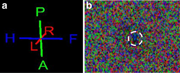
Functional magnetic resonance (fMR) imaging offers plenty of new opportunities in the diagnosis of central nervous system diseases. Diffusion tensor imaging (DTI) is a technique sensitive to the random motion of water providing information about tissue architecture. We applied DTI to normal appearing spinal cords of 13 dogs of different breeds and body weights in a 3.0 T magnetic resonance (MR) scanner. The aim was to study fiber tracking (FT) patterns by tractography and the variations of the fractional anisotropy (FA) and the apparent diffusion coefficient (ADC) observed in the spinal cords of dogs with different sizes and at different locations (cervical and thoracolumbar). For that reason we added a DTI sequence to the standard clinical MR protocol. The values of FA and ADC were calculated by means of three regions of interest defined on the cervical or the thoracolumbar spinal cord (ROI 1, 2, and 3).
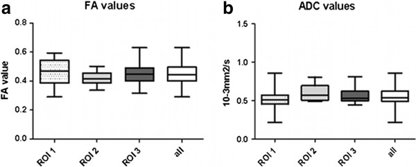
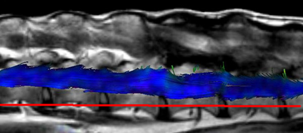
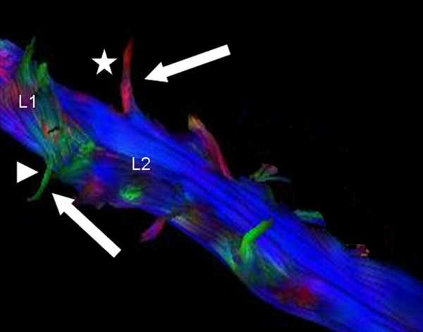
The shape of the spinal cord fiber tracts was well illustrated following tractography and the exiting nerve roots could be differentiated from the spinal cord fiber tracts. Routine MR scanning times were extended for 8 to 12 min, depending on the size of the field of view (FOV), the slice thickness, and the size of the interslice gaps. In small breed dogs (<15 kg body weight) the fibers could be tracked over a length of approximately 10 vertebral bodies with scanning times of about 8 min, whereas in large breed dogs (>25 kg body weight) the traceable fiber length was about 5 vertebral bodies which took 10 to 12 min scanning time. FA and ADC values showed mean values of 0.447 (FA), and 0.560×10(-3) mm2/s (ADC), respectively without any differences detected with regard to different dog sizes and spinal cord 45 segments examined.
FT is suitable for the graphical depiction of the canine spinal cord and the exiting nerve roots. The FA and ADC values offer an objective measure for evaluation of the spinal cord fiber integrity in dogs.
功能磁共振成像为中枢神经系统疾病的诊断提供了大量新机会。扩散张量成像(DTI)是一种对水的随机运动敏感的技术,可提供有关组织结构的信息。我们在一台3.0 T磁共振(MR)扫描仪中,对13只不同品种和体重的犬的外观正常脊髓应用了DTI。目的是通过纤维束成像研究纤维追踪(FT)模式,以及观察不同大小和不同位置(颈段和胸腰段)犬脊髓中各向异性分数(FA)和表观扩散系数(ADC)的变化。因此,我们在标准临床MR协议中增加了一个DTI序列。FA和ADC值通过在颈段或胸腰段脊髓上定义的三个感兴趣区域(ROI 1、2和3)进行计算。
纤维束成像后,脊髓纤维束的形态得到了很好的显示,并且可以将穿出的神经根与脊髓纤维束区分开来。常规MR扫描时间延长了8至12分钟,这取决于视野(FOV)的大小、层厚和层间距。在小型犬(体重<15 kg)中,纤维可以追踪约10个椎体的长度,扫描时间约为8分钟,而在大型犬(体重>25 kg)中,可追踪的纤维长度约为5个椎体,扫描时间为10至12分钟。FA和ADC值的平均值分别为0.447(FA)和0.560×10(-3) mm2/s(ADC),在不同犬只大小和所检查的脊髓节段方面未检测到任何差异。
FT适用于犬脊髓和穿出神经根的图形描绘。FA和ADC值为评估犬脊髓纤维完整性提供了客观指标。