Leibniz-Institute for Age Research, Fritz-Lipman-Institute, Jena, Germany.
PLoS One. 2013 Apr 26;8(4):e62018. doi: 10.1371/journal.pone.0062018. Print 2013.
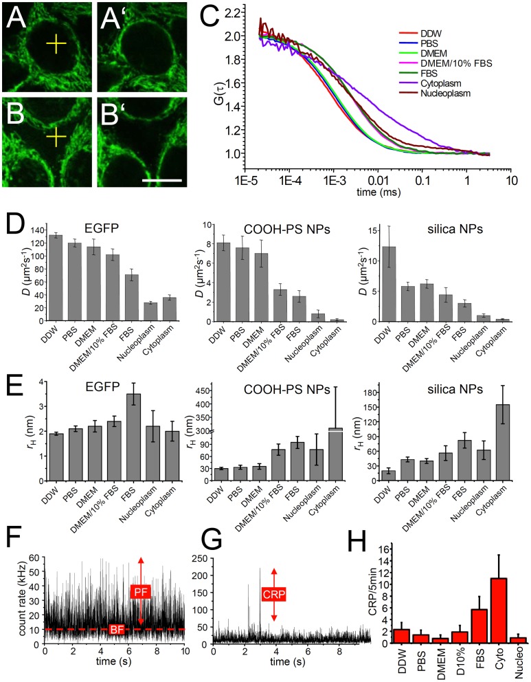
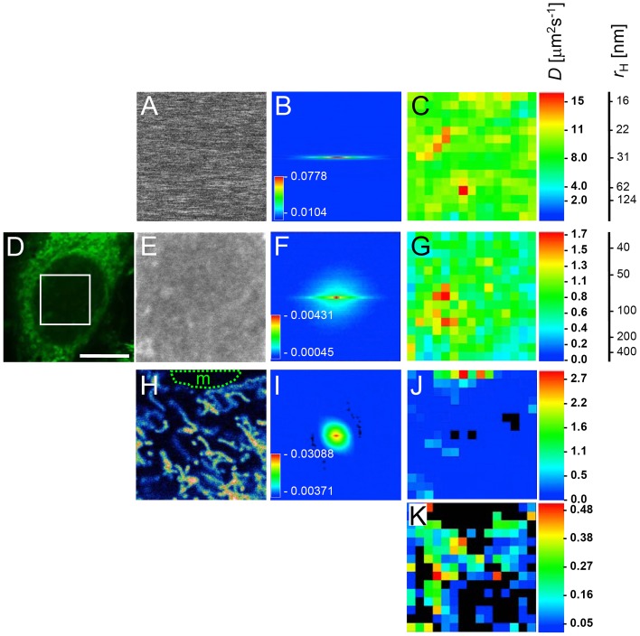
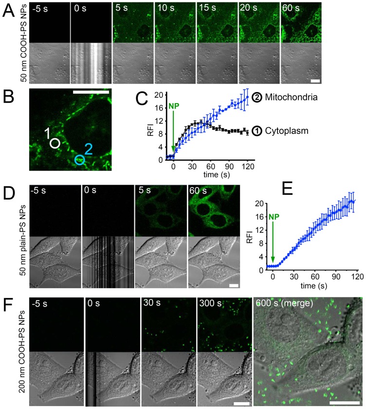
Understanding of nanoparticle-bio-interactions within living cells requires knowledge about the dynamic behavior of nanomaterials during their cellular uptake, intracellular traffic and mutual reactions with cell organelles. Here, we introduce a protocol of combined kinetic imaging techniques that enables investigation of exemplary fluorochrome-labelled nanoparticles concerning their intracellular fate. By time-lapse confocal microscopy we observe fast, dynamin-dependent uptake of polystyrene and silica nanoparticles via the cell membrane within seconds. Fluorescence recovery after photobleaching (FRAP) experiments reveal fast and complete exchange of the investigated nanoparticles at mitochondria, cytoplasmic vesicles or the nuclear envelope. Nuclear translocation is observed within minutes by free diffusion and active transport. Fluorescence correlation spectroscopy (FCS) and raster image correlation spectroscopy (RICS) indicate diffusion coefficients of polystyrene and silica nanoparticles in the nucleus and the cytoplasm that are consistent with particle motion in living cells based on diffusion. Determination of the apparent hydrodynamic radii by FCS and RICS shows that nanoparticles exert their cytoplasmic and nuclear effects mainly as mobile, monodisperse entities. Thus, a complete toolkit of fluorescence fluctuation microscopy is presented for the investigation of nanomaterial biophysics in subcellular microenvironments that contributes to develop a framework of intracellular nanoparticle delivery routes.
要了解纳米颗粒与活细胞内的相互作用,需要了解纳米材料在细胞摄取、细胞内运输以及与细胞器相互作用过程中的动态行为。在这里,我们介绍了一种组合动力学成像技术的方案,该方案可用于研究典型荧光标记纳米颗粒的细胞内命运。通过延时共聚焦显微镜,我们观察到聚苯乙烯和二氧化硅纳米颗粒在几秒钟内通过细胞膜快速、依赖于动力蛋白的摄取。荧光漂白后恢复(FRAP)实验表明,在 线粒体、细胞质囊泡或核膜处,研究的纳米颗粒能快速且完全交换。通过自由扩散和主动运输,在几分钟内观察到核转位。荧光相关光谱(FCS)和光栅图像相关光谱(RICS)表明,聚苯乙烯和二氧化硅纳米颗粒在核和细胞质中的扩散系数与基于扩散的活细胞内颗粒运动一致。通过 FCS 和 RICS 确定的表观流体力学半径表明,纳米颗粒主要作为可移动的单分散体发挥其细胞质和核效应。因此,为研究亚细胞微环境中的纳米材料生物物理学,提出了一套完整的荧光波动显微镜工具,有助于构建细胞内纳米颗粒输送途径的框架。