Center for Medical Physics and Biomedical Engineering, Medical University of Vienna Vienna, Austria ; MR Centre of Excellence, Medical University of Vienna Vienna, Austria ; Department of Statistics and Probability Theory, Vienna University of Technology Vienna, Austria.
Front Hum Neurosci. 2013 May 1;7:168. doi: 10.3389/fnhum.2013.00168. eCollection 2013.
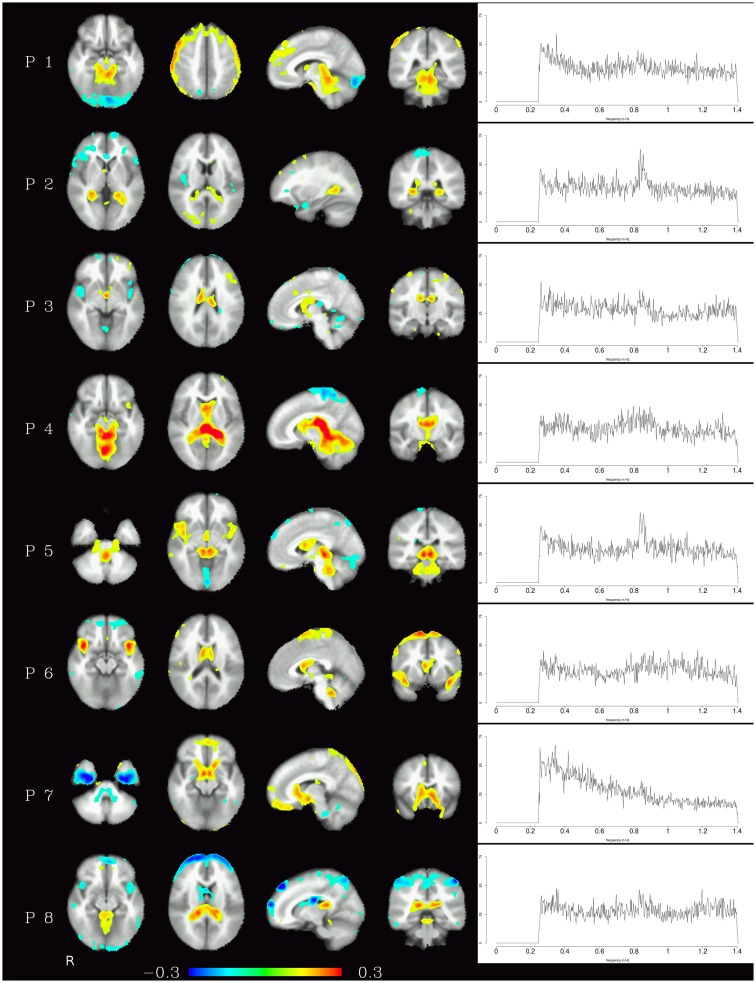
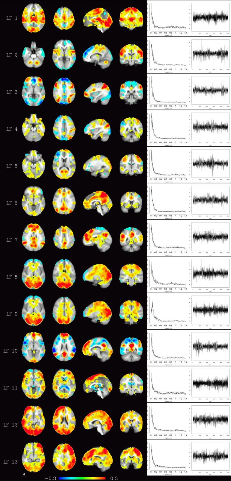
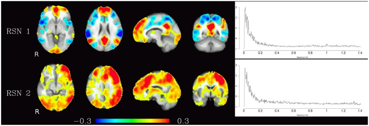
Analysis of resting-state networks using fMRI usually ignores high-frequency fluctuations in the BOLD signal - be it because of low TR prohibiting the analysis of fluctuations with frequencies higher than 0.25 Hz (for a typical TR of 2 s), or because of the application of a bandpass filter (commonly restricting the signal to frequencies lower than 0.1 Hz). While the standard model of convolving neuronal activity with a hemodynamic response function suggests that the signal of interest in fMRI is characterized by slow fluctuation, it is in fact unclear whether the high-frequency dynamics of the signal consists of noise only. In this study, 10 subjects were scanned at 3 T during 6 min of rest using a multiband EPI sequence with a TR of 354 ms to critically sample fluctuations of up to 1.4 Hz. Preprocessed data were high-pass filtered to include only frequencies above 0.25 Hz, and voxelwise whole-brain temporal ICA (tICA) was used to identify consistent high-frequency signals. The resulting components include physiological background signal sources, most notably pulsation and heart-beat components, that can be specifically identified and localized with the method presented here. Perhaps more surprisingly, common resting-state networks like the default-mode network also emerge as separate tICA components. This means that high-frequency oscillations sampled with a rather T1-weighted contrast still contain specific information on these resting-state networks to consistently identify them, not consistent with the commonly held view that these networks operate on low-frequency fluctuations alone. Consequently, the use of bandpass filters in resting-state data analysis should be reconsidered, since this step eliminates potentially relevant information. Instead, more specific methods for the elimination of physiological background signals, for example by regression of physiological noise components, might prove to be viable alternatives.
使用 fMRI 分析静息态网络时,通常会忽略 BOLD 信号中的高频波动——无论是因为 TR 较低而禁止分析频率高于 0.25 Hz 的波动(对于典型的 TR 为 2 s),还是因为应用了带通滤波器(通常将信号限制在低于 0.1 Hz 的频率范围内)。虽然将神经元活动与血液动力学响应函数卷积的标准模型表明 fMRI 中感兴趣的信号的特征是缓慢波动,但实际上尚不清楚信号的高频动态是否仅由噪声组成。在这项研究中,10 名受试者在 3 T 下接受了 6 分钟的休息扫描,使用 TR 为 354 ms 的多频带 EPI 序列进行扫描,以对高达 1.4 Hz 的波动进行关键采样。预处理后的数据进行高通滤波,仅包含高于 0.25 Hz 的频率,并使用全脑时变独立成分分析(tICA)来识别一致的高频信号。由此产生的成分包括生理背景信号源,特别是脉动和心跳成分,可以使用这里提出的方法进行专门识别和定位。也许更令人惊讶的是,像默认模式网络这样的常见静息态网络也作为独立的 tICA 成分出现。这意味着,使用相当 T1 加权对比采样的高频振荡仍然包含有关这些静息态网络的特定信息,以便一致地识别它们,这与这些网络仅在低频波动上运行的普遍观点不一致。因此,在静息态数据分析中使用带通滤波器应该重新考虑,因为这一步骤消除了潜在的相关信息。相反,通过回归生理噪声成分等更具体的方法来消除生理背景信号可能被证明是可行的替代方法。