• 文献检索
  • 文档翻译
  • 深度研究
  • 学术资讯
  • Suppr Zotero 插件Zotero 插件
  • 邀请有礼
  • 套餐&价格
  • 历史记录
应用&插件
Suppr Zotero 插件Zotero 插件浏览器插件Mac 客户端Windows 客户端微信小程序
定价
高级版会员购买积分包购买API积分包
服务
文献检索文档翻译深度研究API 文档MCP 服务
关于我们
关于 Suppr公司介绍联系我们用户协议隐私条款
关注我们

Suppr 超能文献

核心技术专利:CN118964589B侵权必究
粤ICP备2023148730 号-1Suppr @ 2026

文献检索

告别复杂PubMed语法,用中文像聊天一样搜索,搜遍4000万医学文献。AI智能推荐,让科研检索更轻松。

立即免费搜索

文件翻译

保留排版,准确专业,支持PDF/Word/PPT等文件格式,支持 12+语言互译。

免费翻译文档

深度研究

AI帮你快速写综述,25分钟生成高质量综述,智能提取关键信息,辅助科研写作。

立即免费体验

儿童膝关节骺软骨及关节软骨 T1ρ 成像

T1ρ mapping of pediatric epiphyseal and articular cartilage in the knee.

机构信息

Vanderbilt University Department of Biomedical Engineering, Nashville, Tennessee, USA.

出版信息

J Magn Reson Imaging. 2013 Aug;38(2):299-305. doi: 10.1002/jmri.23963. Epub 2013 May 6.

DOI:10.1002/jmri.23963
PMID:23650103
原文链接:https://pmc.ncbi.nlm.nih.gov/articles/PMC3919676/
Abstract

PURPOSE

To evaluate the feasibility of measuring T1ρ values in epiphyseal cartilage in children, we have conducted a novel study of spin locking techniques. Adult articular cartilage has been widely studied with spin locking techniques by magnetic resonance imaging. However, no results are available for in vivo T1ρ imaging of developing cartilage.

MATERIALS AND METHODS

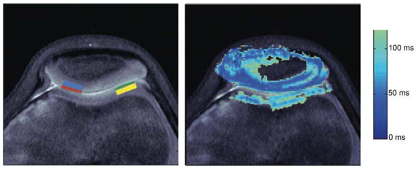
Ten volunteers of age 6 ± 3 years were recruited to have T1ρ mapping performed on the knee at the conclusion of their clinical study. T1ρ maps were generated using a spin-lock cluster followed by a fast spin-echo imaging sequence. Regions of interest (ROIs) were placed in non-load-bearing (NLB), load-bearing (LB), and articular cartilage.

RESULTS

Student's t-tests were performed to compare means among the ROIs. Mean T1ρ for epiphyseal and articular cartilage was 49.8 ± 9 and 76.6 ± 7 ms, respectively. LB and NLB T1ρ vales were 47.1 ± 9.5 and 52.5 ± 9 ms, respectively. Significant differences were found in T1ρ values between epiphyseal and articular cartilage layers (P = 0.0001). No difference in T1ρ was observed between NLB and LB layers. A modest trend was also noted for epiphyseal and articular cartilage regions with age.

CONCLUSION

It is feasible to quantify differences in epiphyseal and articular cartilage layers with SL techniques. T1ρ holds promise as a noninvasive method of studying normal and abnormal developmental states of cartilage in children.

摘要

目的

为了评估在儿童骺软骨中测量 T1ρ 值的可行性,我们进行了一项新的自旋锁定技术研究。磁共振成像已广泛研究了成人关节软骨的自旋锁定技术,但尚无关于发育中软骨的体内 T1ρ 成像的结果。

材料和方法

招募了 10 名年龄为 6 ± 3 岁的志愿者,在完成临床研究后对膝关节进行 T1ρ 图谱分析。T1ρ 图谱是通过自旋锁定簇后快速自旋回波成像序列生成的。感兴趣区(ROI)放置在非承重区(NLB)、承重区(LB)和关节软骨区。

结果

对 ROI 之间的平均值进行了学生 t 检验。骺软骨和关节软骨的平均 T1ρ 值分别为 49.8 ± 9ms 和 76.6 ± 7ms。LB 和 NLB 的 T1ρ 值分别为 47.1 ± 9.5ms 和 52.5 ± 9ms。骺软骨和关节软骨层之间的 T1ρ 值存在显著差异(P = 0.0001)。NLB 和 LB 层之间的 T1ρ 值无差异。骺软骨和关节软骨区域与年龄也存在适度的趋势。

结论

SL 技术可以定量分析骺软骨和关节软骨层之间的差异。T1ρ 有望成为一种非侵入性方法,用于研究儿童软骨的正常和异常发育状态。

https://cdn.ncbi.nlm.nih.gov/pmc/blobs/76d2/3919676/1d6d21410328/nihms520770f5.jpg
https://cdn.ncbi.nlm.nih.gov/pmc/blobs/76d2/3919676/59cf0cb78560/nihms520770f1.jpg
https://cdn.ncbi.nlm.nih.gov/pmc/blobs/76d2/3919676/a2bc9498812b/nihms520770f2.jpg
https://cdn.ncbi.nlm.nih.gov/pmc/blobs/76d2/3919676/0e3e2e52385a/nihms520770f3.jpg
https://cdn.ncbi.nlm.nih.gov/pmc/blobs/76d2/3919676/c95f6fcd4484/nihms520770f4.jpg
https://cdn.ncbi.nlm.nih.gov/pmc/blobs/76d2/3919676/1d6d21410328/nihms520770f5.jpg

相似文献

1
T1ρ mapping of pediatric epiphyseal and articular cartilage in the knee.儿童膝关节骺软骨及关节软骨 T1ρ 成像
J Magn Reson Imaging. 2013 Aug;38(2):299-305. doi: 10.1002/jmri.23963. Epub 2013 May 6.
2
Rapid 3D-T1rho mapping of the knee joint at 3.0T with parallel imaging.3.0T下膝关节的快速3D-T1rho成像及并行成像技术
Magn Reson Med. 2006 Sep;56(3):563-71. doi: 10.1002/mrm.20982.
3
Validation and optimization of adiabatic T and T for quantitative imaging of articular cartilage at 3 T.3T 下用于关节软骨定量成像的绝热 T 和 T 的验证与优化
Magn Reson Med. 2017 Mar;77(3):1265-1275. doi: 10.1002/mrm.26183. Epub 2016 Mar 4.
4
Repeatability of Quantitative Knee Cartilage T, T, and T Mapping With 3D-MRI Fingerprinting.三维磁共振指纹技术定量膝关节软骨 T1、T2 和 T2* 值的可重复性。
J Magn Reson Imaging. 2024 Aug;60(2):688-699. doi: 10.1002/jmri.29068. Epub 2023 Oct 26.
5
3D-T1rho quantitation of patellar cartilage at 3.0T.3.0T下髌骨软骨的3D-T1rho定量分析
J Magn Reson Imaging. 2006 Dec;24(6):1357-63. doi: 10.1002/jmri.20733.
6
Relationship between knee alignment and T1ρ values of articular cartilage and menisci in patients with knee osteoarthritis.膝关节骨关节炎患者膝关节对线与关节软骨和半月板 T1ρ 值的关系。
Eur J Radiol. 2013 Nov;82(11):1946-52. doi: 10.1016/j.ejrad.2013.05.010. Epub 2013 Jun 13.
7
High resolution T1ρ mapping of in vivo human knee cartilage at 7T.7T下人体膝关节软骨的高分辨率T1ρ成像
PLoS One. 2014 May 15;9(5):e97486. doi: 10.1371/journal.pone.0097486. eCollection 2014.
8
Simultaneous acquisition of T1ρ and T2 quantification in knee cartilage: repeatability and diurnal variation.膝关节软骨中T1ρ与T2定量的同步采集:重复性与日变化
J Magn Reson Imaging. 2014 May;39(5):1287-93. doi: 10.1002/jmri.24253. Epub 2013 Jul 29.
9
Quantitative T2 and T1ρ mapping are sensitive to ischemic injury to the epiphyseal cartilage in an in vivo piglet model of Legg-Calvé-Perthes disease.定量 T2 和 T1ρ 图谱对活体小猪莱格-卡尔弗-佩尔特斯病模型中骺软骨的缺血性损伤敏感。
Osteoarthritis Cartilage. 2022 Sep;30(9):1244-1253. doi: 10.1016/j.joca.2022.05.009. Epub 2022 May 26.
10
Quantitative evaluation of knee cartilage and meniscus destruction in patients with rheumatoid arthritis using T1ρ and T2 mapping.使用 T1ρ 和 T2 映射定量评估类风湿关节炎患者的膝关节软骨和半月板破坏。
Eur J Radiol. 2017 Nov;96:91-97. doi: 10.1016/j.ejrad.2017.09.018. Epub 2017 Oct 3.

引用本文的文献

1
Normal subchondral high T2 signal on MRI mimicking sacroiliitis in children: frequency, age distribution, and relationship to skeletal maturity.MRI 上正常的软骨下高 T2 信号模拟儿童的骶髂关节炎:频率、年龄分布与骨骼成熟度的关系。
Eur Radiol. 2021 May;31(5):3498-3507. doi: 10.1007/s00330-020-07328-0. Epub 2020 Oct 29.
2
T-mapping for assessing knee joint cartilage in children with juvenile idiopathic arthritis - feasibility and repeatability.T 映射评估儿童幼年特发性关节炎膝关节软骨 - 可行性和可重复性。
Pediatr Radiol. 2020 Mar;50(3):371-379. doi: 10.1007/s00247-019-04557-4. Epub 2019 Nov 9.
3
Diagnostic value of T1ρ and T2 mapping sequences of 3D fat-suppressed spoiled gradient (FS SPGR-3D) 3.0-T magnetic resonance imaging for osteoarthritis.3.0-T磁共振成像3D脂肪抑制扰相梯度回波(FS SPGR-3D)序列的T1ρ和T2映射对骨关节炎的诊断价值
Medicine (Baltimore). 2019 Jan;98(1):e13834. doi: 10.1097/MD.0000000000013834.
4
New insights into rotating frame relaxation at high field.高场下旋转坐标系弛豫的新见解。
NMR Biomed. 2016 Sep;29(9):1258-73. doi: 10.1002/nbm.3490. Epub 2016 Feb 11.
5
Capturing fast relaxing spins with SWIFT adiabatic rotating frame spin-lattice relaxation (T1ρ) mapping.利用SWIFT绝热旋转框架自旋晶格弛豫(T1ρ)映射捕获快速弛豫自旋。
NMR Biomed. 2016 Apr;29(4):420-30. doi: 10.1002/nbm.3474. Epub 2016 Jan 26.
6
Chemical exchange in knee cartilage assessed by R1ρ (1/T1ρ) dispersion at 3T.通过3T下的R1ρ(1/T1ρ)色散评估膝关节软骨中的化学交换。
Magn Reson Imaging. 2015 Jan;33(1):38-42. doi: 10.1016/j.mri.2014.07.008. Epub 2014 Aug 2.

本文引用的文献

1
Chemical exchange saturation transfer (CEST): what is in a name and what isn't?化学交换饱和传递(CEST):名字里有什么,又没有什么?
Magn Reson Med. 2011 Apr;65(4):927-48. doi: 10.1002/mrm.22761. Epub 2011 Feb 17.
2
The effects of acute loading on T1rho and T2 relaxation times of tibiofemoral articular cartilage.急性加载对胫股关节软骨 T1rho 和 T2 弛豫时间的影响。
Osteoarthritis Cartilage. 2010 Dec;18(12):1557-63. doi: 10.1016/j.joca.2010.10.001. Epub 2010 Oct 13.
3
MR imaging insights into skeletal maturation: what is normal?磁共振成像对骨骼成熟的见解:什么是正常情况?
Radiology. 2009 Jan;250(1):28-38. doi: 10.1148/radiol.2501071322.
4
Assessment of glycosaminoglycan concentration in vivo by chemical exchange-dependent saturation transfer (gagCEST).通过化学交换依赖饱和转移(gagCEST)对体内糖胺聚糖浓度进行评估。
Proc Natl Acad Sci U S A. 2008 Feb 19;105(7):2266-70. doi: 10.1073/pnas.0707666105. Epub 2008 Feb 11.
5
Three-dimensional delayed gadolinium-enhanced MRI of cartilage (dGEMRIC) for in vivo evaluation of reparative cartilage after matrix-associated autologous chondrocyte transplantation at 3.0T: Preliminary results.3.0T磁共振成像下三维延迟钆增强磁共振成像(dGEMRIC)用于体内评估基质相关自体软骨细胞移植后修复软骨的初步结果
J Magn Reson Imaging. 2007 Oct;26(4):974-82. doi: 10.1002/jmri.21091.
6
2007 Elizabeth Winston Lanier Award Winner. Magnetic resonance imaging of cartilage glycosaminoglycan: basic principles, imaging technique, and clinical applications.2007年伊丽莎白·温斯顿·拉尼尔奖获得者。软骨糖胺聚糖的磁共振成像:基本原理、成像技术及临床应用。
J Orthop Res. 2008 Mar;26(3):281-91. doi: 10.1002/jor.20482.
7
The effect of varying echo spacing within a multiecho acquisition: better characterization of long T2 components.多回波采集中不同回波间隔的影响:对长T2成分的更好表征
Magn Reson Imaging. 2007 Jul;25(6):840-7. doi: 10.1016/j.mri.2006.09.046. Epub 2007 Apr 5.
8
In vivo T(1rho) and T(2) mapping of articular cartilage in osteoarthritis of the knee using 3 T MRI.使用3T磁共振成像对膝关节骨关节炎患者的关节软骨进行体内T(1rho)和T(2)成像
Osteoarthritis Cartilage. 2007 Jul;15(7):789-97. doi: 10.1016/j.joca.2007.01.011. Epub 2007 Feb 16.
9
Artifacts in T1 rho-weighted imaging: compensation for B(1) and B(0) field imperfections.T1ρ加权成像中的伪影:B(1)和B(0)场缺陷的补偿
J Magn Reson. 2007 May;186(1):75-85. doi: 10.1016/j.jmr.2007.01.015. Epub 2007 Jan 26.
10
Early ischemia in growing piglet skeleton: MR diffusion and perfusion imaging.生长中小猪骨骼的早期缺血:磁共振扩散和灌注成像
Radiology. 2007 Jan;242(1):129-36. doi: 10.1148/radiol.2421050680.