Department of Neurology, Max Planck Institute for Human Cognitive and Brain Sciences Leipzig, Germany ; Clinic for Cognitive Neurology, University of Leipzig Leipzig, Germany.
Front Hum Neurosci. 2013 May 7;7:183. doi: 10.3389/fnhum.2013.00183. eCollection 2013.
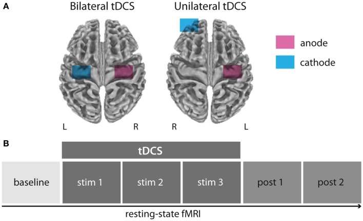
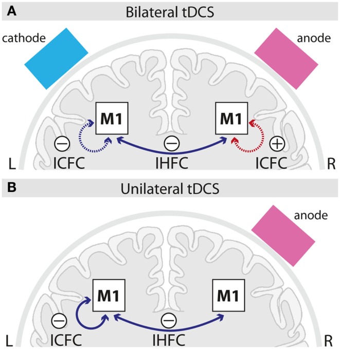
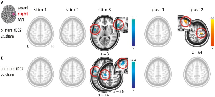
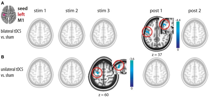
Transcranial direct current stimulation (tDCS) over the primary motor cortex (M1) has been shown to induce changes in motor performance and learning. Recent studies indicate that tDCS is capable of modulating widespread neural network properties within the brain. However the temporal evolution of online- and after-effects of tDCS on functional connectivity (FC) within and across the stimulated motor cortices (M1) still remain elusive. In the present study, two different tDCS setups were investigated: (i) unilateral M1 tDCS (anode over right M1, cathode over the contralateral supraorbital region) and (ii) bilateral M1 tDCS (anode over right M1, cathode over left M1). In a randomized single-blinded cross-over design, 12 healthy subjects underwent functional magnetic resonance imaging at rest before, during and after 20 min of either bi-, unilateral, or sham M1 tDCS. Seed-based FC analysis was used to investigate tDCS-induced changes across and within M1. We found that bilateral M1 tDCS induced (a) a decrease in interhemispheric FC during stimulation and (b) an increase in intracortical FC within right M1 after termination of the intervention. While unilateral M1 tDCS also resulted in similar effects during stimulation, no such changes could be observed after termination of tDCS. Our results provide evidence that depending on the electrode montage, tDCS acts upon a modulation of either intracortical and/or interhemispheric processing of M1.
经颅直流电刺激(tDCS)作用于初级运动皮层(M1)已被证明可以改变运动表现和学习。最近的研究表明,tDCS 能够调节大脑内广泛的神经网络特性。然而,tDCS 对刺激运动皮层(M1)内和跨皮质的功能连接(FC)的在线和后效的时间演变仍然难以捉摸。在本研究中,我们研究了两种不同的 tDCS 设置:(i)单侧 M1 tDCS(阳极置于右侧 M1,阴极置于对侧眶上区域)和(ii)双侧 M1 tDCS(阳极置于右侧 M1,阴极置于左侧 M1)。在随机单盲交叉设计中,12 名健康受试者在接受 20 分钟双侧、单侧或假 M1 tDCS 之前、期间和之后进行了静息状态下的功能磁共振成像。基于种子的 FC 分析用于研究 M1 内和跨 M1 的 tDCS 诱导变化。我们发现,双侧 M1 tDCS 诱导(a)刺激期间半球间 FC 降低,(b)干预结束后右侧 M1 内皮质内 FC 增加。虽然单侧 M1 tDCS 在刺激期间也产生了类似的效果,但在 tDCS 结束后观察不到这种变化。我们的结果提供了证据,表明根据电极排列方式,tDCS 作用于 M1 的皮质内和/或半球间处理的调制。