Department of Health Care, Qilu Hospital of Shandong University, Jinan, China.
PLoS One. 2013 May 13;8(5):e62723. doi: 10.1371/journal.pone.0062723. Print 2013.
To analyze the diagnostic value of color Doppler imaging (CDI) of blood flow in the retrobulbar vessels of eyes with primary open-angle glaucoma (POAG).
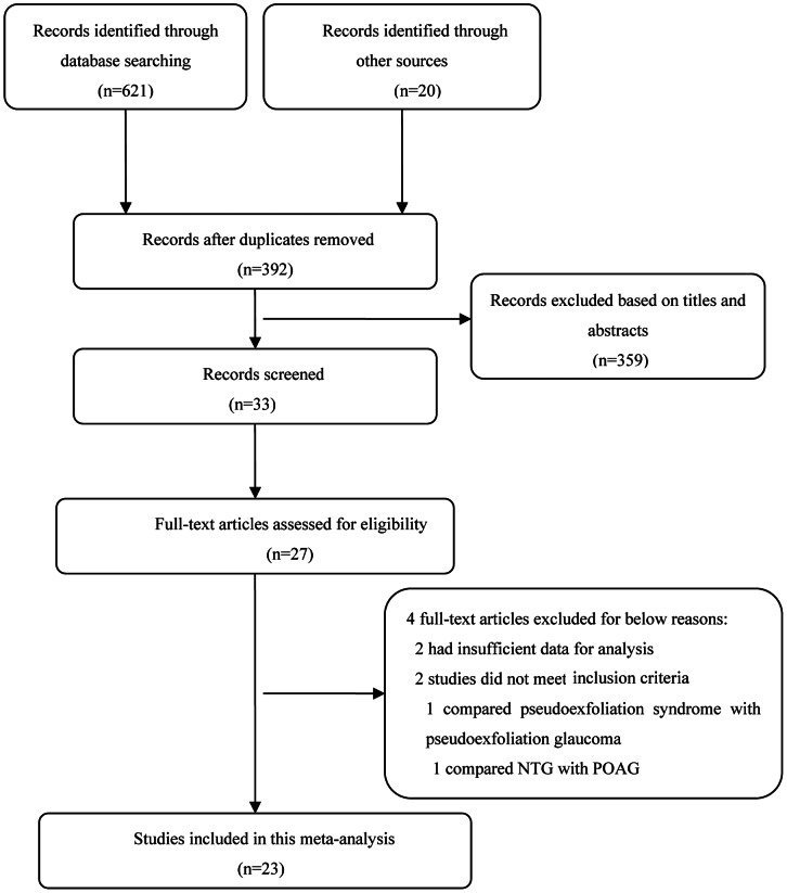
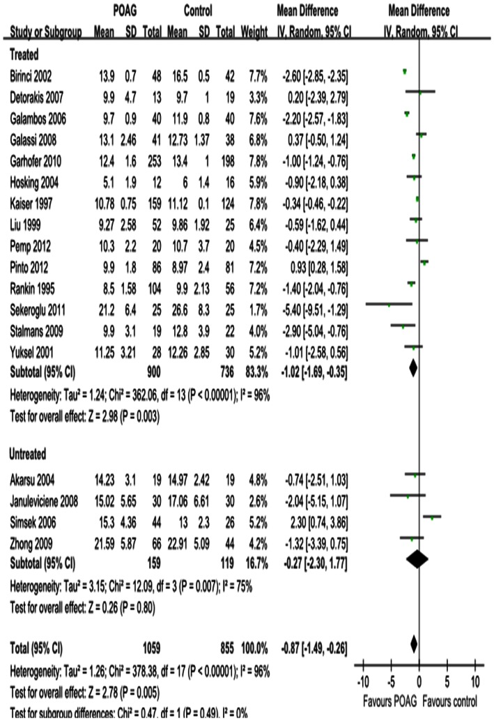
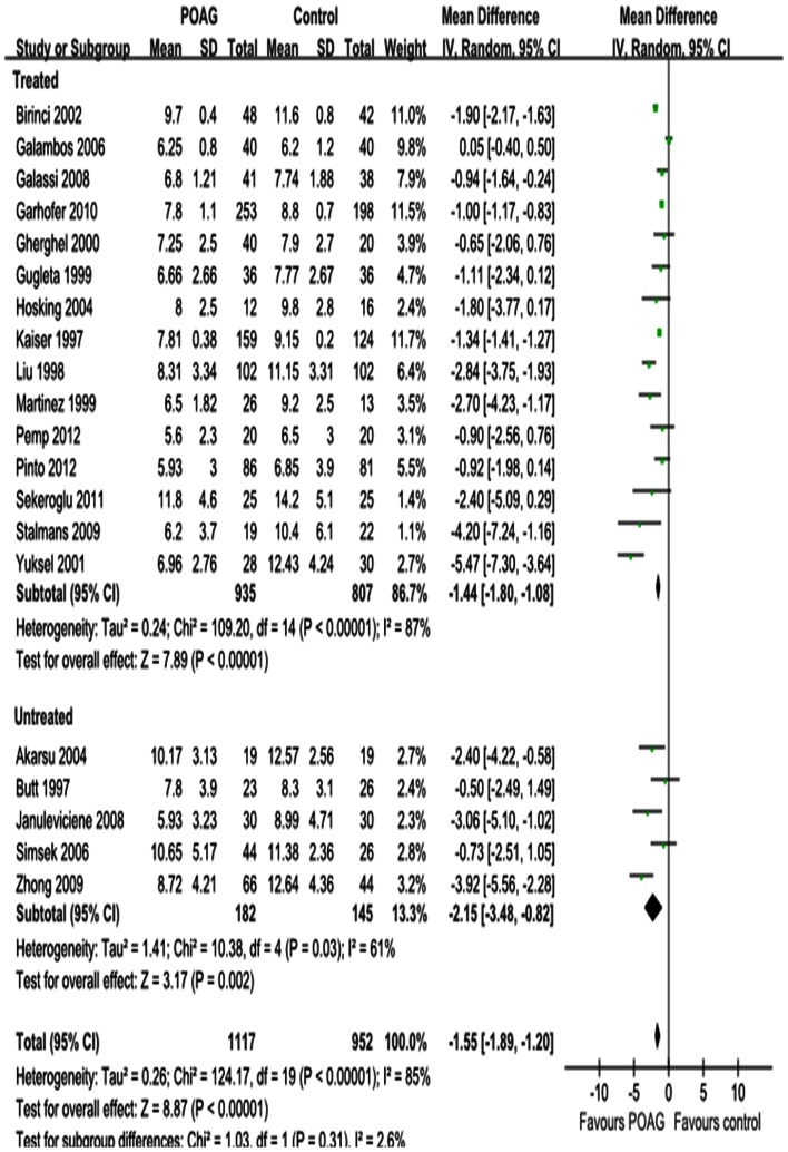
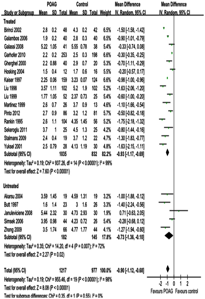
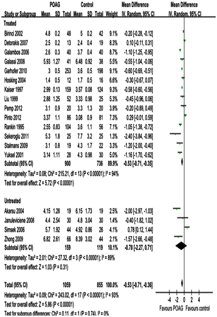
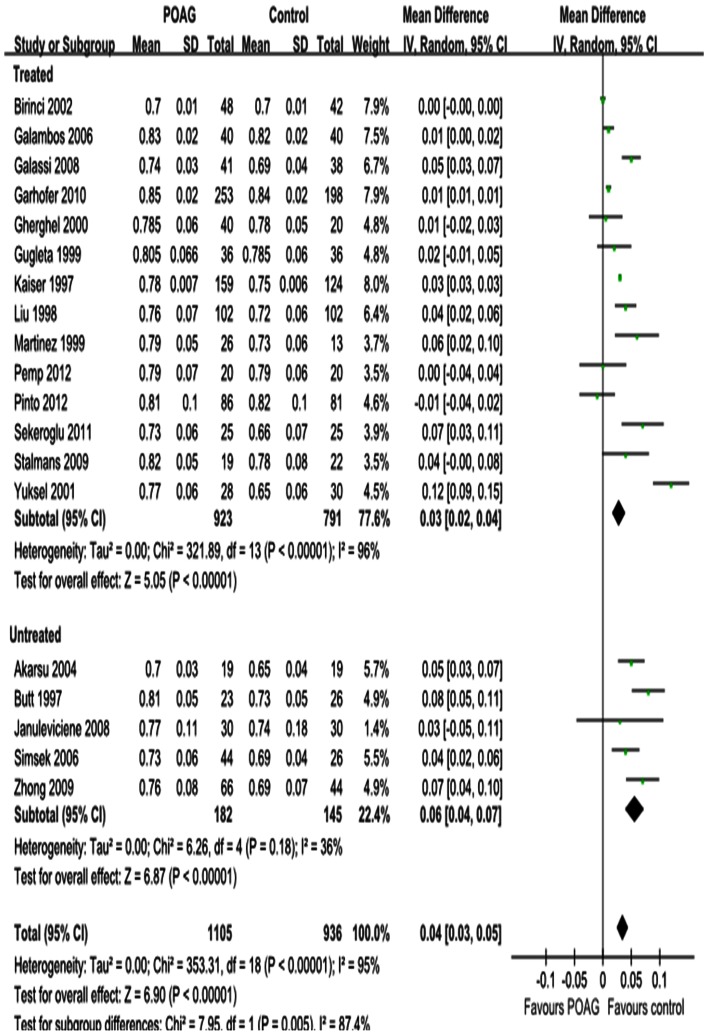
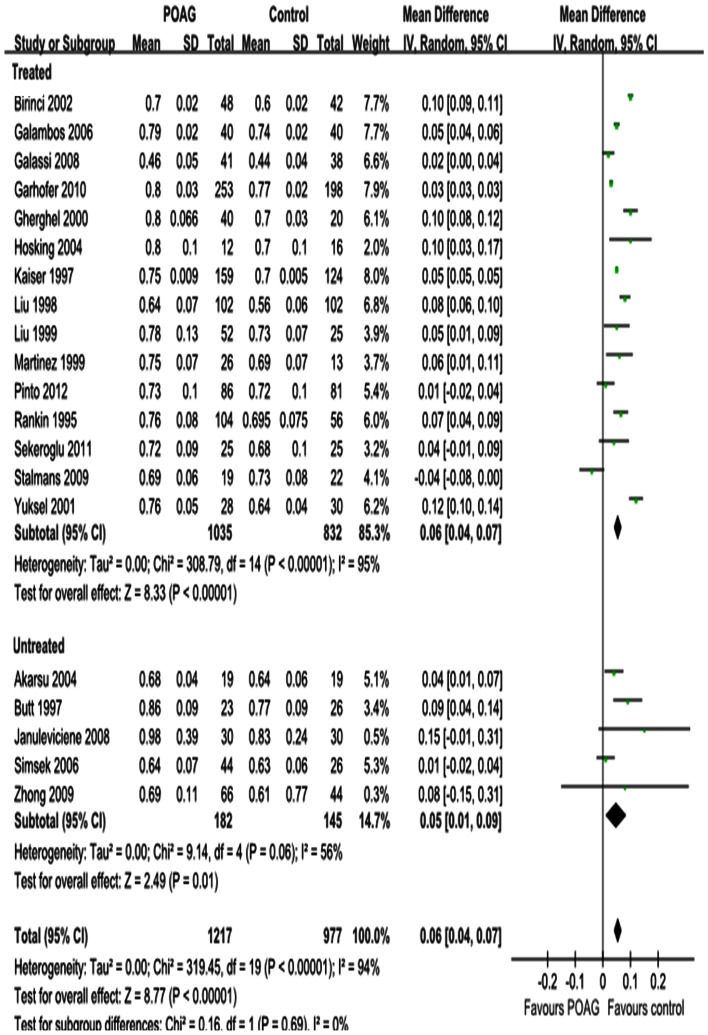
Pertinent publications were retrieved from the Cochrane Central Register of Controlled Trials, PubMed and the ISI Web of Knowledge up to October 2012. Changes in peak systolic velocity (PSV), end diastolic velocity (EDV) and resistive index (RI) of the ophthalmic artery (OA), central retinal artery (CRA) and short posterior ciliary artery (SPCA) of POAG eyes and normal controls were evaluated by CDI. Subgroup analyses were conducted according to whether patients received IOP-lowering drugs treatment and were defined as treated and untreated.
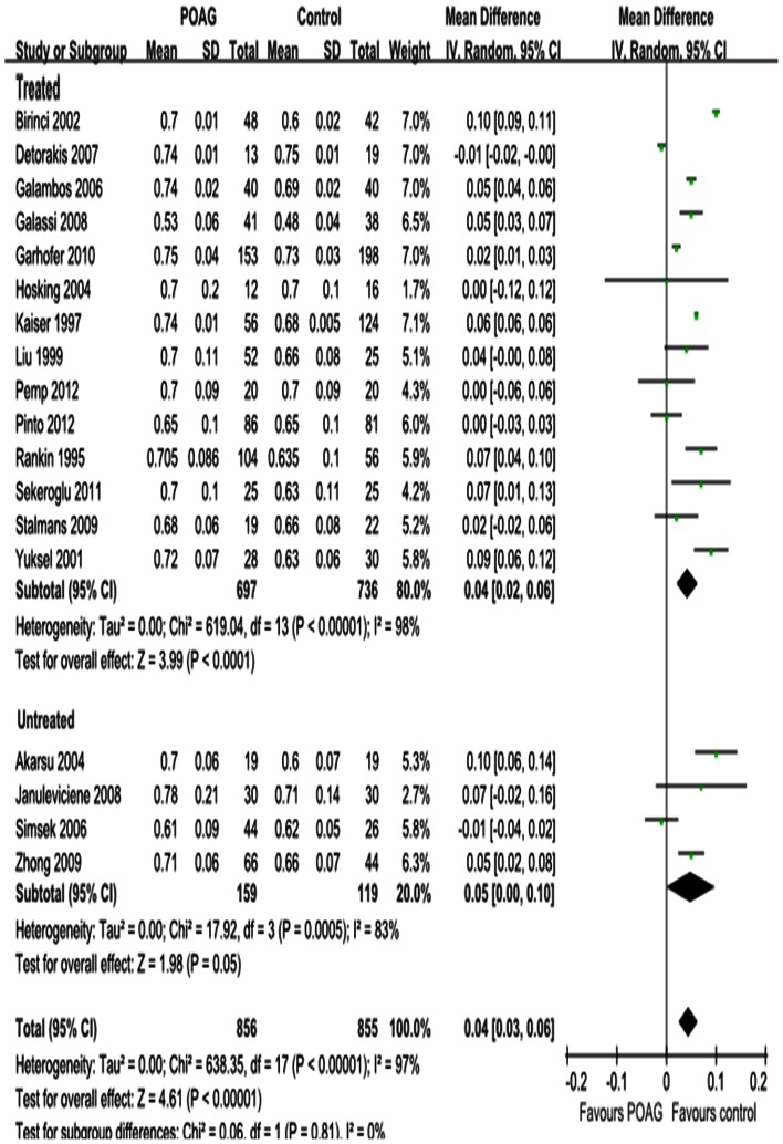
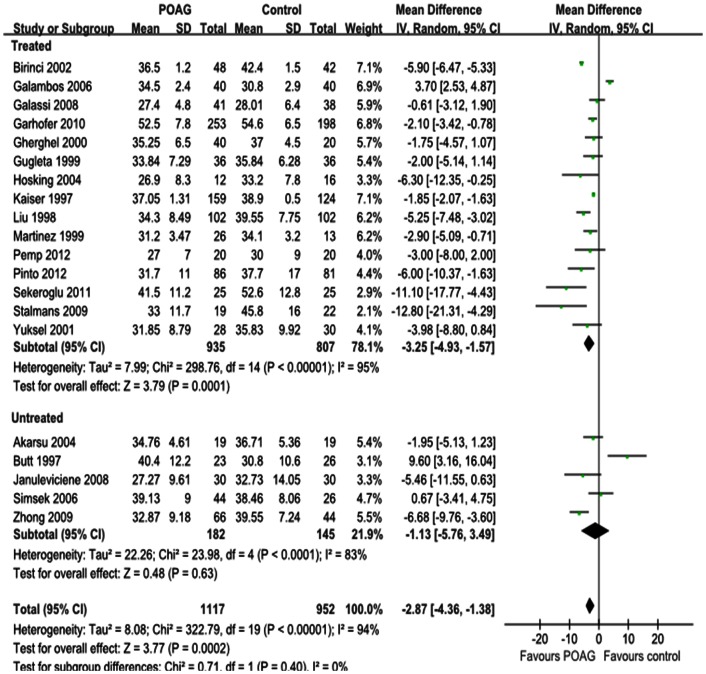
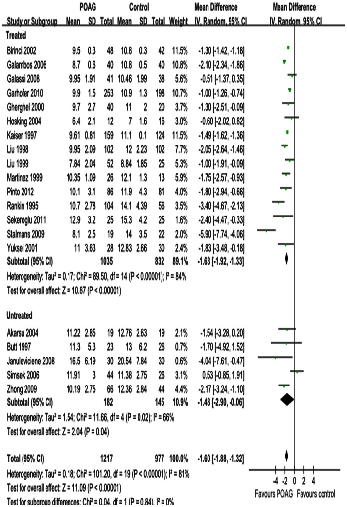
PSV and EDV were statistically significantly reduced in the OA of POAG eyes (P = 0.0002; P<0.00001; respectively), with significant heterogeneity (P(heterogeneity)<0.00001, I² = 94%; P(heterogeneity)<0.00001, I² = 85%; respectively). Similar results were demonstrated for the CRA (P<0.00001; respectively) and SPCA (P = 0.005; P<0.00001; respectively), with significant heterogeneities for both the CRA (P(heterogeneity)<0.00001, I² = 81%; P(heterogeneity)<0.00001, I² = 98%; respectively) and the SPCA (P(heterogeneity)<0.00001, I² = 96%; P(heterogeneity)<0.00001, I² = 93%; respectively). Significant increases in RI were found in all retrobulbar vessels (P<0.00001; respectively), with significant heterogeneities (P(heterogeneity)<0.00001, I² = 95%; P(heterogeneity)<0.00001, I² = 94%; P(heterogeneity)<0.00001, I² = 97%; respectively).
This meta-analysis suggests that CDI is a potential diagnostic tool for POAG.
分析彩色多谱勒成像(CDI)在原发性开角型青光眼(POAG)患者眼动静脉血流中的诊断价值。
从 Cochrane 对照试验中心注册库、PubMed 和 ISI Web of Knowledge 中检索截至 2012 年 10 月的相关文献。通过 CDI 评估 POAG 眼和正常对照组眼的眼动脉(OA)、视网膜中央动脉(CRA)和睫状后短动脉(SPCA)的收缩期峰值速度(PSV)、舒张末期速度(EDV)和阻力指数(RI)的变化。根据患者是否接受降眼压药物治疗将患者分为治疗组和未治疗组进行亚组分析。
POAG 眼的 OA 的 PSV 和 EDV 显著降低(P=0.0002;P<0.00001;分别),且具有显著的异质性(P(异质性)<0.00001,I²=94%;P(异质性)<0.00001,I²=85%;分别)。CRA(P<0.00001;分别)和 SPCA(P=0.005;P<0.00001;分别)也得到了相似的结果,并且这两条动脉都具有显著的异质性(CRA:P(异质性)<0.00001,I²=81%;P(异质性)<0.00001,I²=98%;分别;SPCA:P(异质性)<0.00001,I²=96%;P(异质性)<0.00001,I²=93%;分别)。所有眼后段血管的 RI 均显著升高(P<0.00001;分别),且具有显著的异质性(P(异质性)<0.00001,I²=95%;P(异质性)<0.00001,I²=94%;P(异质性)<0.00001,I²=97%;分别)。
这项荟萃分析表明,CDI 可能是 POAG 的一种潜在诊断工具。