• 文献检索
  • 文档翻译
  • 深度研究
  • 学术资讯
  • Suppr Zotero 插件Zotero 插件
  • 邀请有礼
  • 套餐&价格
  • 历史记录
应用&插件
Suppr Zotero 插件Zotero 插件浏览器插件Mac 客户端Windows 客户端微信小程序
定价
高级版会员购买积分包购买API积分包
服务
文献检索文档翻译深度研究API 文档MCP 服务
关于我们
关于 Suppr公司介绍联系我们用户协议隐私条款
关注我们

Suppr 超能文献

核心技术专利:CN118964589B侵权必究
粤ICP备2023148730 号-1Suppr @ 2026

文献检索

告别复杂PubMed语法,用中文像聊天一样搜索,搜遍4000万医学文献。AI智能推荐,让科研检索更轻松。

立即免费搜索

文件翻译

保留排版,准确专业,支持PDF/Word/PPT等文件格式,支持 12+语言互译。

免费翻译文档

深度研究

AI帮你快速写综述,25分钟生成高质量综述,智能提取关键信息,辅助科研写作。

立即免费体验

将眼血流生物标志物与正常眼压性青光眼疑似患者的筛板参数相关联。与青光眼患者和健康对照者进行比较。

Associating the biomarkers of ocular blood flow with lamina cribrosa parameters in normotensive glaucoma suspects. Comparison to glaucoma patients and healthy controls.

机构信息

Department of Ophthalmology, Wroclaw Medical University, Wroclaw, Poland.

Department of Biomedical Engineering, Faculty of Fundamental Problems of Technology, Wroclaw University of Science and Technology, Wroclaw, Poland.

出版信息

PLoS One. 2021 Mar 23;16(3):e0248851. doi: 10.1371/journal.pone.0248851. eCollection 2021.

DOI:10.1371/journal.pone.0248851
PMID:33755663
原文链接:https://pmc.ncbi.nlm.nih.gov/articles/PMC7987161/
Abstract

PURPOSE

To evaluate association between ocular blood flow biomarkers and lamina cribrosa parameters in normotensive glaucoma suspects compared to glaucoma patients and healthy controls.

METHODS

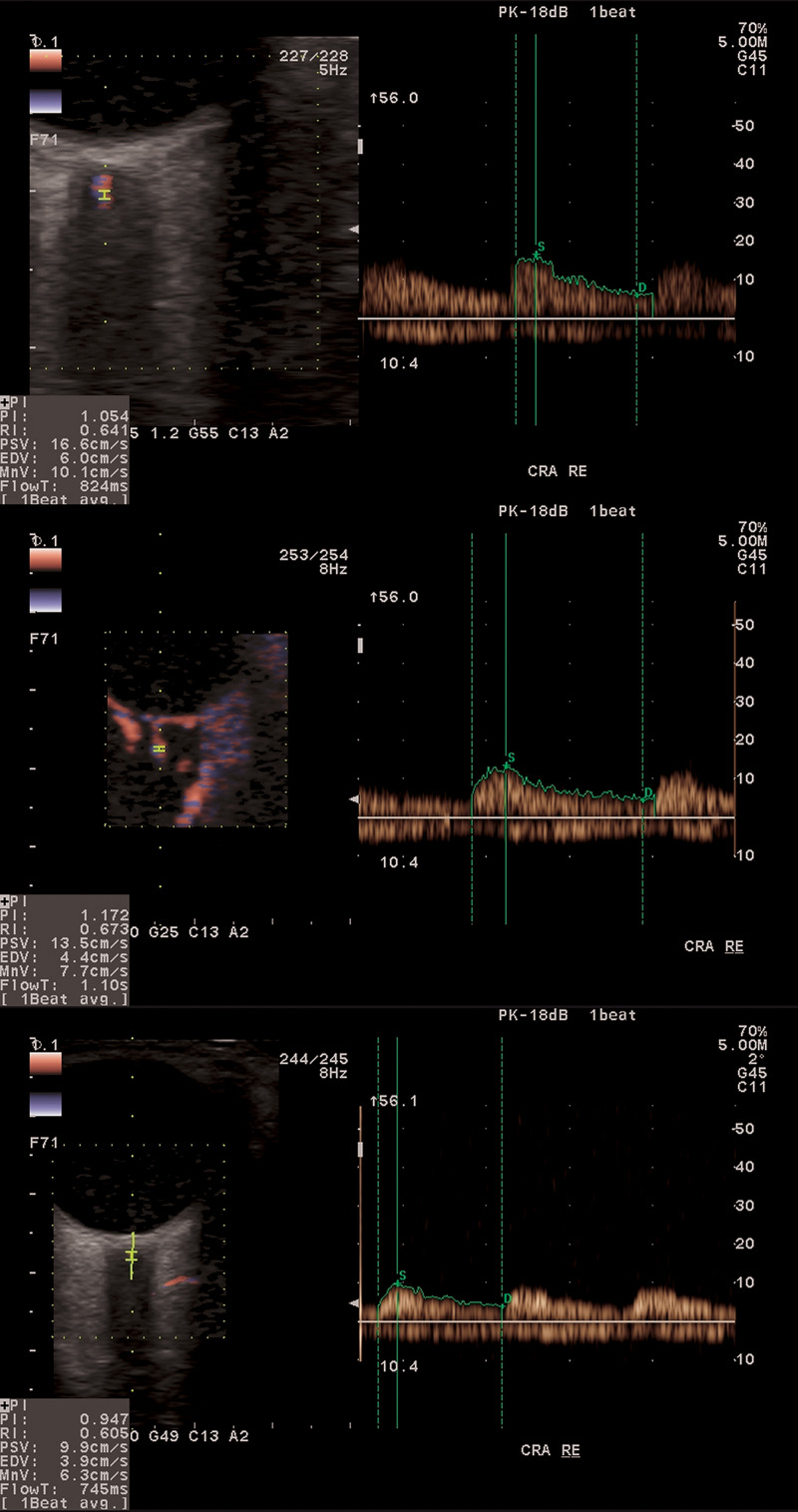
A total of 211 subjects (72 normotensive glaucoma suspects, 70 with primary open-angle glaucoma and 69 controls) were included. Ocular blood flow biomarkers in ophthalmic artery, central retinal artery, as well as in nasal and temporal short posterior ciliary arteries were measured using colour Doppler imaging. Lamina cribrosa position was assessed by measuring its depth, deflection depth, lamina cribrosa shape index and its horizontal equivalent (LCSIH) on B-scan images obtained using optical coherence tomography.

RESULTS

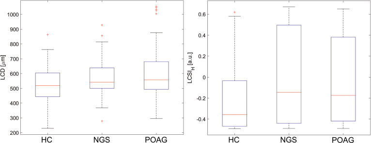
Ocular blood flow biomarkers in glaucoma patients were statistically significantly reduced when compared to healthy controls in peak systolic velocity (PSV) (P = 0.001 in ophthalmic artery and P<0.001 in central retinal artery) and mean flow velocity (Vm) (P = 0.008 in ophthalmic artery and P = 0.008 in central retinal artery), but not statistically significantly different to that of glaucoma suspects except for PSV in central retinal artery (P = 0.011). Statistically significant correlations corrected for age, central corneal thickness and intraocular pressure were found in glaucoma patients between LCSIH and end diastolic velocity of central retinal artery (P = 0.011), and of nasal short posterior ciliary artery (P = 0.028), and between LCSIH and Vm of central retinal artery (P = 0.011) and of nasal short posterior ciliary artery (P = 0.007). No significant correlations were observed between these parameters in glaucoma suspects and healthy controls.

CONCLUSIONS

Impaired ocular blood flow associated with the deformation of lamina cribrosa was found in glaucoma patients, whereas glaucoma suspects had similar lamina cribrosa shape to glaucoma patients but that deformation was not associated with ocular blood flow biomarkers.

摘要

目的

评估正常眼压性青光眼患者与开角型青光眼患者和健康对照者的眼血流生物标志物与筛板参数之间的相关性。

方法

共纳入 211 名受试者(72 名正常眼压性青光眼患者、70 名开角型青光眼患者和 69 名健康对照者)。使用彩色多普勒成像测量眼动脉、视网膜中央动脉以及鼻侧和颞侧短睫状后动脉的血流生物标志物。使用光学相干断层扫描获得 B 扫描图像,评估筛板位置,测量其深度、凹陷深度、筛板形状指数及其水平等效值(LCSIH)。

结果

与健康对照组相比,青光眼患者的眼血流生物标志物在收缩期峰值速度(PSV)(眼动脉 P = 0.001,视网膜中央动脉 P<0.001)和平均流速(Vm)(眼动脉 P = 0.008,视网膜中央动脉 P = 0.008)方面均显著降低,但与青光眼患者无统计学差异,除视网膜中央动脉 PSV 外(P = 0.011)。在青光眼患者中,LCSIH 与视网膜中央动脉的舒张末期速度(P = 0.011)和鼻侧短睫状后动脉(P = 0.028),以及与视网膜中央动脉的 Vm(P = 0.011)和鼻侧短睫状后动脉(P = 0.007)之间存在统计学显著相关性,校正年龄、中央角膜厚度和眼内压后相关性仍具有统计学意义。在青光眼患者和健康对照者中,这些参数之间均无显著相关性。

结论

在青光眼患者中发现了与筛板变形相关的眼部血流受损,而青光眼患者的筛板形状与青光眼患者相似,但这种变形与眼部血流生物标志物无关。

https://cdn.ncbi.nlm.nih.gov/pmc/blobs/de7c/7987161/fcd47839d9d5/pone.0248851.g002.jpg
https://cdn.ncbi.nlm.nih.gov/pmc/blobs/de7c/7987161/f93b4258627b/pone.0248851.g001.jpg
https://cdn.ncbi.nlm.nih.gov/pmc/blobs/de7c/7987161/fcd47839d9d5/pone.0248851.g002.jpg
https://cdn.ncbi.nlm.nih.gov/pmc/blobs/de7c/7987161/f93b4258627b/pone.0248851.g001.jpg
https://cdn.ncbi.nlm.nih.gov/pmc/blobs/de7c/7987161/fcd47839d9d5/pone.0248851.g002.jpg

相似文献

1
Associating the biomarkers of ocular blood flow with lamina cribrosa parameters in normotensive glaucoma suspects. Comparison to glaucoma patients and healthy controls.将眼血流生物标志物与正常眼压性青光眼疑似患者的筛板参数相关联。与青光眼患者和健康对照者进行比较。
PLoS One. 2021 Mar 23;16(3):e0248851. doi: 10.1371/journal.pone.0248851. eCollection 2021.
2
Lamina Cribrosa Depth and Shape in Glaucoma Suspects. Comparison to Glaucoma Patients and Healthy Controls.青光眼疑似患者的神经纤维层筛板深度和形状。与青光眼患者和健康对照者的比较。
Curr Eye Res. 2019 Sep;44(9):1026-1033. doi: 10.1080/02713683.2019.1616767. Epub 2019 May 23.
3
Ocular hemodynamics in pseudoexfoliation syndrome and pseudoexfoliation glaucoma.假性剥脱综合征和假性剥脱性青光眼中的眼部血流动力学
Ophthalmology. 2001 Jun;108(6):1043-9. doi: 10.1016/s0161-6420(01)00572-3.
4
Relationship between ocular perfusion pressure and retrobulbar blood flow in patients with glaucoma with progressive damage.青光眼病情进展性损害患者的眼灌注压与球后血流之间的关系
Am J Ophthalmol. 2000 Nov;130(5):597-605. doi: 10.1016/s0002-9394(00)00766-2.
5
Correlation among choroidal, parapapillary, and retrobulbar vascular parameters in glaucoma.青光眼患者脉络膜、视乳头旁及球后血管参数之间的相关性
Am J Ophthalmol. 2009 Apr;147(4):736-743.e2. doi: 10.1016/j.ajo.2008.10.020. Epub 2009 Feb 1.
6
Trabeculectomy is associated with retrobulbar hemodynamic changes. A color Doppler analysis.小梁切除术与球后血流动力学变化有关。彩色多普勒分析。
Ophthalmology. 1994 Feb;101(2):340-51. doi: 10.1016/s0161-6420(13)31332-3.
7
Ocular blood flow in glaucoma - the Leuven Eye Study.青光眼患者的眼部血流——鲁汶眼研究
Acta Ophthalmol. 2016 Sep;94(6):592-8. doi: 10.1111/aos.12962. Epub 2016 Feb 19.
8
Color Doppler imaging discloses reduced ocular blood flow velocities in nonexudative age-related macular degeneration.彩色多普勒成像显示非渗出性年龄相关性黄斑变性患者眼血流速度降低。
Am J Ophthalmol. 1999 Jul;128(1):75-80. doi: 10.1016/s0002-9394(99)00061-6.
9
Ocular blood flow in healthy and primary open-angle glaucomatous eyes.健康眼和原发性开角型青光眼眼中的眼血流。
Ophthalmologica. 2002 Nov-Dec;216(6):434-7. doi: 10.1159/000067553.
10
Blood-flow velocities of the extraocular vessels in patients with high-tension and normal-tension primary open-angle glaucoma.高眼压和正常眼压原发性开角型青光眼患者眼外血管的血流速度
Am J Ophthalmol. 1997 Mar;123(3):320-7. doi: 10.1016/s0002-9394(14)70127-8.

引用本文的文献

1
The Robust Lamina Cribrosa Vasculature: Perfusion and Oxygenation Under Elevated Intraocular Pressure.稳健的神经节细胞层血管:高眼压下的灌注和氧合。
Invest Ophthalmol Vis Sci. 2024 May 1;65(5):1. doi: 10.1167/iovs.65.5.1.
2
Temporal Protrusion of the Optic Disc Implicated in Central Visual Field Abnormalities in High Myopia: Two Case Reports.高度近视中视盘颞侧突出与中心视野异常相关:两例报告
Case Rep Ophthalmol. 2024 Apr 15;15(1):335-342. doi: 10.1159/000538440. eCollection 2024 Jan-Dec.
3
Unveiling the silent link: Normal-tension glaucoma's enigmatic bond with cardiac blood flow.

本文引用的文献

1
Lamina Cribrosa Depth and Shape in Glaucoma Suspects. Comparison to Glaucoma Patients and Healthy Controls.青光眼疑似患者的神经纤维层筛板深度和形状。与青光眼患者和健康对照者的比较。
Curr Eye Res. 2019 Sep;44(9):1026-1033. doi: 10.1080/02713683.2019.1616767. Epub 2019 May 23.
2
Ocular blood flow in glaucoma - the Leuven Eye Study.青光眼患者的眼部血流——鲁汶眼研究
Acta Ophthalmol. 2016 Sep;94(6):592-8. doi: 10.1111/aos.12962. Epub 2016 Feb 19.
3
Emerging risk factors for glaucoma onset and progression.青光眼发病和进展的新兴危险因素。
揭示沉默的关联:正常眼压性青光眼与心脏血流的神秘联系。
World J Cardiol. 2024 Jan 26;16(1):10-15. doi: 10.4330/wjc.v16.i1.10.
4
Current State of Knowledge in Ocular Blood Flow in Glaucoma: A Narrative Review.青光眼患者眼血流的当前知识状态:一篇叙述性综述。
Clin Ophthalmol. 2023 Aug 31;17:2599-2607. doi: 10.2147/OPTH.S426709. eCollection 2023.
5
The Role of Color Doppler Imaging in the Diagnosis of Glaucoma: A Review of the Literature.彩色多普勒成像在青光眼诊断中的作用:文献综述
Diagnostics (Basel). 2023 Feb 5;13(4):588. doi: 10.3390/diagnostics13040588.
6
Cardiac Hypertrophy May Be a Risk Factor for the Development and Severity of Glaucoma.心脏肥大可能是青光眼发生和严重程度的一个风险因素。
Biomedicines. 2022 Mar 15;10(3):677. doi: 10.3390/biomedicines10030677.
Prog Brain Res. 2015;221:81-101. doi: 10.1016/bs.pbr.2015.04.007. Epub 2015 Jul 2.
4
Age-Related Changes in Ocular Blood Velocity in Suspects with Glaucomatous Optic Disc Appearance. Comparison with Healthy Subjects and Glaucoma Patients.具有青光眼性视盘外观的疑似患者眼部血流速度的年龄相关性变化。与健康受试者和青光眼患者的比较。
PLoS One. 2015 Jul 28;10(7):e0134357. doi: 10.1371/journal.pone.0134357. eCollection 2015.
5
The role of retrobulbar and retinal circulation on optic nerve head and retinal nerve fibre layer structure in patients with open-angle glaucoma over an 18-month period.18个月期间球后及视网膜循环对开角型青光眼患者视神经乳头和视网膜神经纤维层结构的作用。
Br J Ophthalmol. 2015 May;99(5):609-12. doi: 10.1136/bjophthalmol-2014-305780. Epub 2014 Dec 2.
6
Parameters characterizing age-dependent retrobulbar circulation in healthy subjects measured by color Doppler ultrasonography.通过彩色多普勒超声测量的健康受试者中表征年龄依赖性球后循环的参数。
Curr Eye Res. 2015 Jul;40(7):729-36. doi: 10.3109/02713683.2014.954675. Epub 2014 Sep 8.
7
Optic nerve head blood flow response to reduced ocular perfusion pressure by alteration of either the blood pressure or intraocular pressure.通过改变血压或眼压,视神经乳头血流对降低的眼灌注压作出反应。
Curr Eye Res. 2015 Apr;40(4):359-67. doi: 10.3109/02713683.2014.924146. Epub 2014 Jun 9.
8
Higher optic nerve sheath diameters are associated with lower ocular blood flow velocities in glaucoma patients.高的视神经鞘直径与青光眼患者较低的眼血流速度相关。
Graefes Arch Clin Exp Ophthalmol. 2014 Mar;252(3):477-83. doi: 10.1007/s00417-013-2533-y. Epub 2013 Nov 27.
9
Ocular perfusion pressure in glaucoma.青光眼的眼内灌注压。
Acta Ophthalmol. 2014 Jun;92(4):e252-66. doi: 10.1111/aos.12298. Epub 2013 Nov 18.
10
Color Doppler imaging analysis of retrobulbar blood flow velocities in primary open-angle glaucomatous eyes: a meta-analysis.彩色多普勒成像分析原发性开角型青光眼患者眼后血流速度:荟萃分析。
PLoS One. 2013 May 13;8(5):e62723. doi: 10.1371/journal.pone.0062723. Print 2013.