Radin M J, Eaton K A, Krakowka S, Morgan D R, Lee A, Otto G, Fox J
Ohio State University, Columbus 43210.
Infect Immun. 1990 Aug;58(8):2606-12. doi: 10.1128/iai.58.8.2606-2612.1990.
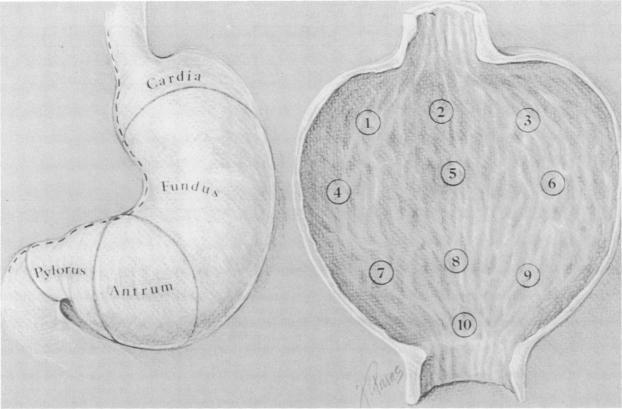
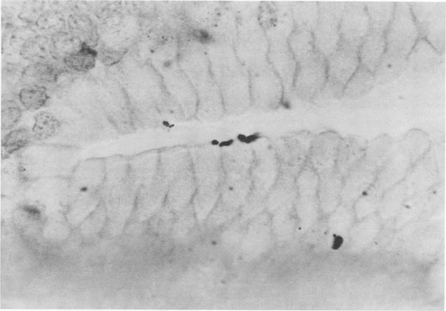
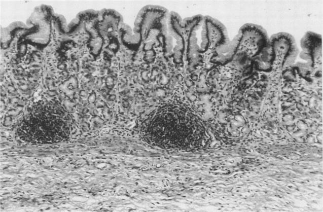
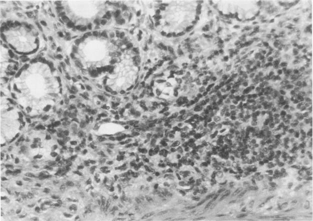
Establishment of infection with Helicobacter pylori and gastritis in nonhuman species is currently only successful in gnotobiotic piglets. This study was designed to determine whether H. pylori will colonize the gastrointestinal tract of gnotobiotic dogs. Gnotobiotic beagle pups were derived by standard methods. Group A (five dogs) was orally challenged with 3 x 10(8) H. pylori at 7 days of age. Group B (two dogs) received only peptone water but was contact-exposed beginning on day 23 postinfection (p.i.). Necropsy was performed on dogs on day 30 p.i. H. pylori colonized the stomach of all dogs (groups A and B). Urease map analysis correlated with the microbiologic findings and indicated that the density of colonization was less than that observed in human tissue. Organisms were also recovered from the pharynx, esophagus, duodenum, and rectum of 1, 2, 2, and 1 dog, respectively. All group A and one group B dog developed serum immunoglobulin G specific for H. pylori by day 30 p.i. Gross lesions were restricted to the stomach and consisted of small (less than 1 mm) lymphoid follicles. Microscopically, there were focal to diffuse lymphoplasmacytic infiltrates with follicle formation and mild to moderate infiltration of neutrophils and eosinophils in the gastric lamina propria. With the Warthin-Starry silver stain, organisms were seen on the surface of the gastric epithelial cells, beneath the mucus layer. We conclude that H. pylori colonizes the stomachs of gnotobiotic dogs for at least 1 month and the lesions resemble those seen in humans. H. pylori is transmissible by contact from infected to noninfected dogs.
目前,仅在无菌仔猪中成功建立了幽门螺杆菌感染及胃炎模型。本研究旨在确定幽门螺杆菌是否会在无菌犬的胃肠道中定植。无菌比格幼犬通过标准方法获得。A组(5只犬)在7日龄时经口用3×10⁸幽门螺杆菌攻击。B组(2只犬)仅接受蛋白胨水,但在感染后第23天开始接触感染。在感染后第30天对犬进行尸检。幽门螺杆菌定植于所有犬(A组和B组)的胃中。尿素酶图谱分析与微生物学结果相关,表明定植密度低于在人体组织中观察到的密度。分别从1只、2只、2只和1只犬的咽部、食管、十二指肠和直肠中也分离出了细菌。到感染后第30天,所有A组犬和1只B组犬都产生了针对幽门螺杆菌的血清免疫球蛋白G。大体病变仅限于胃,表现为小(小于1毫米)的淋巴滤泡。显微镜下,胃固有层有局灶性至弥漫性的淋巴浆细胞浸润并伴有滤泡形成,以及中性粒细胞和嗜酸性粒细胞的轻度至中度浸润。用Warthin-Starry银染色法,可在胃上皮细胞表面、黏液层下方看到细菌。我们得出结论,幽门螺杆菌可在无菌犬的胃中定植至少1个月,且病变与人类所见相似。幽门螺杆菌可通过接触从感染犬传播至未感染犬。