Hashimoto Yasuhiro, Okamoto Akiko, Saitoh Hisao, Hatakeyama Shingo, Yoneyama Takahiro, Koie Takuya, Ohyama Chikara
Oyokyo Kidney Research Institute, Hirosaki Hospital, Hirosaki 036-8243, Japan ; Department of Urology, Hirosaki University Graduate School of Medicine, 5 Zaifucho, Hirosaki, Aomori 036-8562, Japan.
Int J Nephrol. 2013;2013:980923. doi: 10.1155/2013/980923. Epub 2013 Apr 27.
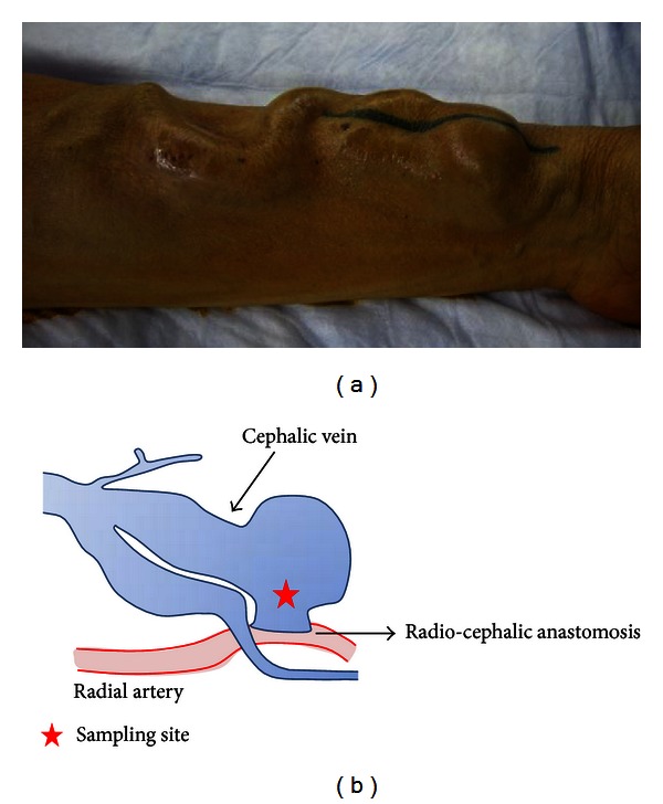
Aim. The objective of this study was to characterize coordinated molecular changes in the structure and composition of the walls of venous segments of arteriovenous (AV) fistulas evoked by overflow. Methods. Venous tissue samples were collected from 6 hemodialysis patients with AV fistulas exposed to overflow and from the normal cephalic veins of 4 other hemodialysis patients. Total RNA was extracted from the venous tissue samples, and gene expression between the 2 groups was compared using Whole Human Genome DNA microarray 44 K. Microarray data were analyzed by GeneSpring GX software and Ingenuity Pathway Analysis. Results. The cDNA microarray analysis identified 397 upregulated genes and 456 downregulated genes. Gene ontology analysis with GeneSpring GX software revealed that biological developmental processes and glycosaminoglycan binding were the most upregulated. In addition, most upregulation occurred extracellularly. In the pathway analysis, the TGF beta signaling pathway, cytokines and inflammatory response pathway, hypertrophy model, and the myometrial relaxation and contraction pathway were significantly upregulated compared with the control cephalic vein. Conclusion. Combining microarray results and pathway information available via the Internet provided biological insight into the structure and composition of the venous wall of overflow AV fistulas.
目的。本研究的目的是描述由血流过载引起的动静脉(AV)内瘘静脉段壁结构和组成的协调性分子变化。方法。从6例暴露于血流过载的AV内瘘血液透析患者以及另外4例血液透析患者的正常头静脉采集静脉组织样本。从静脉组织样本中提取总RNA,并使用全人类基因组DNA微阵列44K比较两组之间的基因表达。微阵列数据通过GeneSpring GX软件和Ingenuity通路分析进行分析。结果。cDNA微阵列分析鉴定出397个上调基因和456个下调基因。使用GeneSpring GX软件进行的基因本体分析显示,生物发育过程和糖胺聚糖结合上调最为明显。此外,大多数上调发生在细胞外。在通路分析中,与对照头静脉相比,转化生长因子β信号通路、细胞因子和炎症反应通路、肥大模型以及子宫肌层舒张和收缩通路显著上调。结论。结合微阵列结果和通过互联网获得的通路信息,为血流过载AV内瘘静脉壁的结构和组成提供了生物学见解。