• 文献检索
  • 文档翻译
  • 深度研究
  • 学术资讯
  • Suppr Zotero 插件Zotero 插件
  • 邀请有礼
  • 套餐&价格
  • 历史记录
应用&插件
Suppr Zotero 插件Zotero 插件浏览器插件Mac 客户端Windows 客户端微信小程序
定价
高级版会员购买积分包购买API积分包
服务
文献检索文档翻译深度研究API 文档MCP 服务
关于我们
关于 Suppr公司介绍联系我们用户协议隐私条款
关注我们

Suppr 超能文献

核心技术专利:CN118964589B侵权必究
粤ICP备2023148730 号-1Suppr @ 2026

文献检索

告别复杂PubMed语法,用中文像聊天一样搜索,搜遍4000万医学文献。AI智能推荐,让科研检索更轻松。

立即免费搜索

文件翻译

保留排版,准确专业,支持PDF/Word/PPT等文件格式,支持 12+语言互译。

免费翻译文档

深度研究

AI帮你快速写综述,25分钟生成高质量综述,智能提取关键信息,辅助科研写作。

立即免费体验

脊椎动物脑生发层中 prominin-1(CD133)阳性细胞的空间分布。

Spatial distribution of prominin-1 (CD133)-positive cells within germinative zones of the vertebrate brain.

机构信息

Tissue Engineering Laboratories-BIOTEC, Medical Faculty Carl Gustav Carus, Technische Universität Dresden, Dresden, Germany.

出版信息

PLoS One. 2013 May 27;8(5):e63457. doi: 10.1371/journal.pone.0063457. Print 2013.

DOI:10.1371/journal.pone.0063457
PMID:23723983
原文链接:https://pmc.ncbi.nlm.nih.gov/articles/PMC3664558/
Abstract

BACKGROUND

In mammals, embryonic neural progenitors as well as adult neural stem cells can be prospectively isolated based on the cell surface expression of prominin-1 (CD133), a plasma membrane glycoprotein. In contrast, characterization of neural progenitors in non-mammalian vertebrates endowed with significant constitutive neurogenesis and inherent self-repair ability is hampered by the lack of suitable cell surface markers. Here, we have investigated whether prominin-1-orthologues of the major non-mammalian vertebrate model organisms show any degree of conservation as for their association with neurogenic geminative zones within the central nervous system (CNS) as they do in mammals or associated with activated neural progenitors during provoked neurogenesis in the regenerating CNS.

METHODS

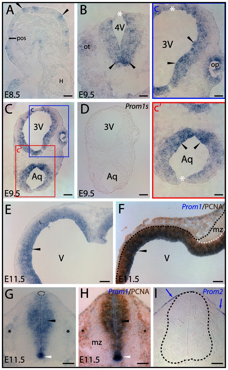
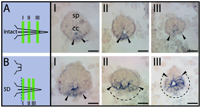
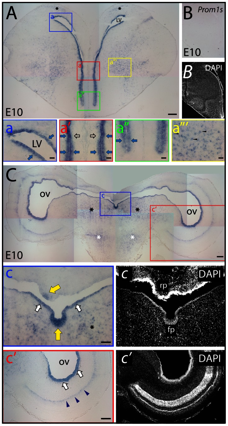
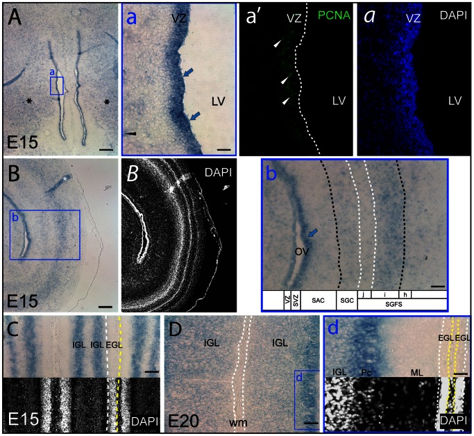
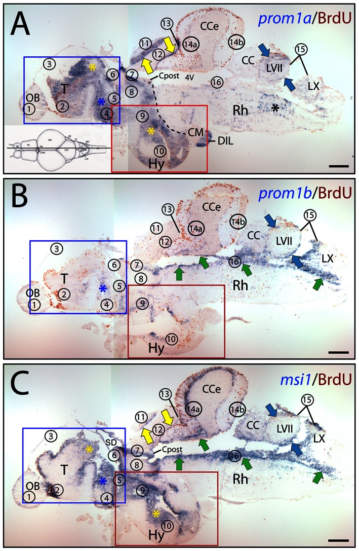
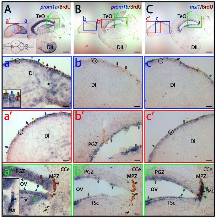
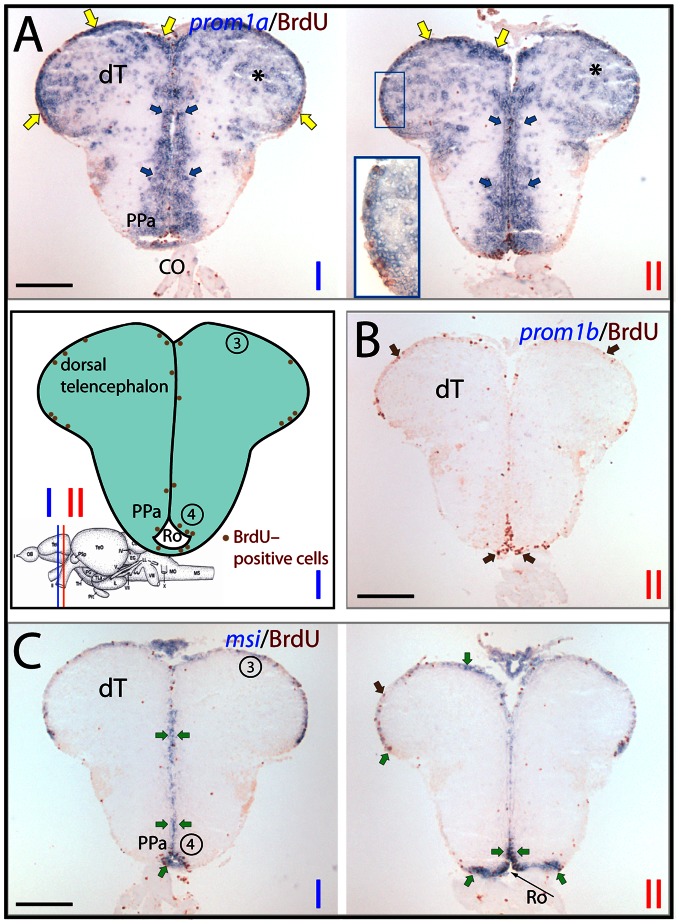
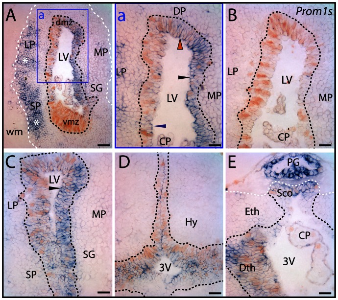
We have recently identified prominin-1 orthologues from zebrafish, axolotl and chicken. The spatial distribution of prominin-1-positive cells--in comparison to those of mice--was mapped in the intact brain in these organisms by non-radioactive in situ hybridization combined with detection of proliferating neural progenitors, marked either by proliferating cell nuclear antigen or 5-bromo-deoxyuridine. Furthermore, distribution of prominin-1 transcripts was investigated in the regenerating spinal cord of injured axolotl.

RESULTS

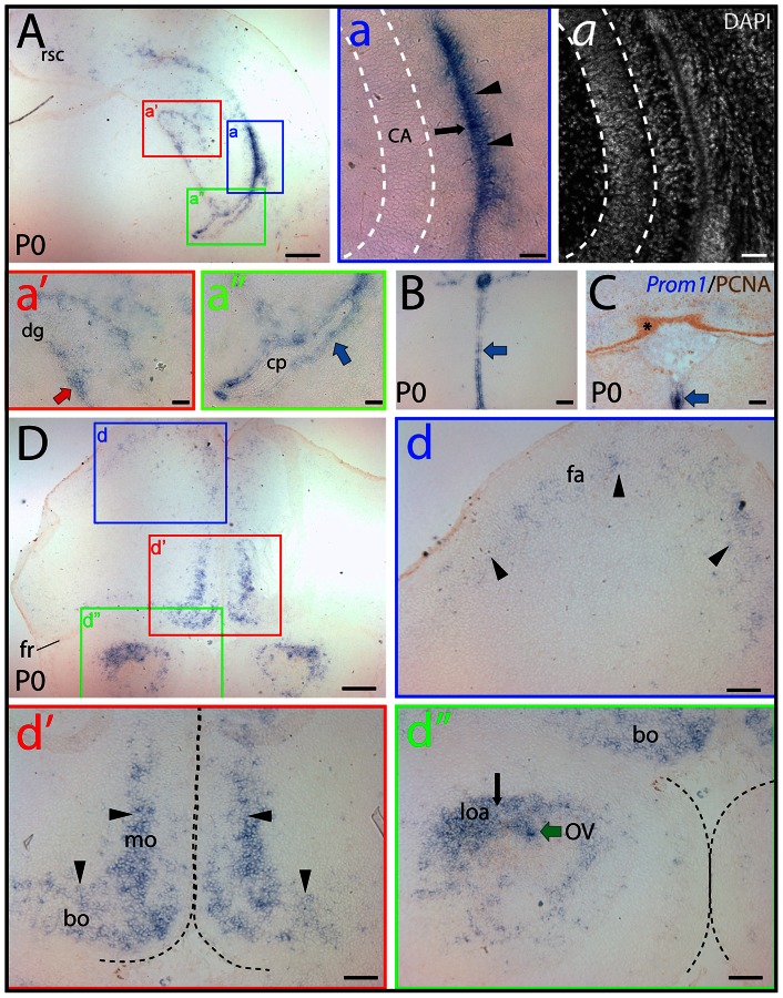
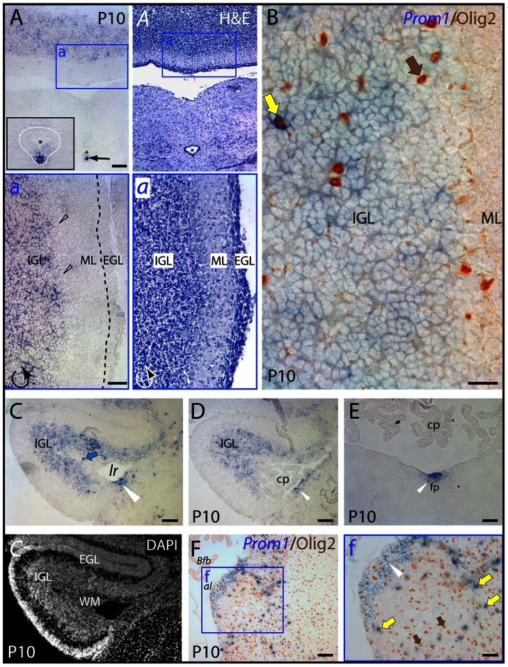
Remarkably, a conserved association of prominin-1 with germinative zones of the CNS was uncovered as manifested in a significant co-localization with cell proliferation markers during normal constitutive neurogenesis in all species investigated. Moreover, an enhanced expression of prominin-1 became evident associated with provoked, compensatory neurogenesis during the epimorphic regeneration of the axolotl spinal cord. Interestingly, significant prominin-1-expressing cell populations were also detected at distinct extraventricular (parenchymal) locations in the CNS of all vertebrate species being suggestive of further, non-neurogenic neural function(s).

CONCLUSION/INTERPRETATION: Collectively, our work provides the first data set describing a comparative analysis of prominin-1-positive progenitor cells across species establishing a framework for further functional characterization in the context of regeneration.

摘要

背景

在哺乳动物中,胚胎神经祖细胞以及成年神经干细胞可以根据质膜糖蛋白 prominin-1(CD133)的细胞表面表达来进行前瞻性分离。相比之下,由于缺乏合适的细胞表面标记物,对具有显著组成性神经发生和固有自我修复能力的非哺乳动物脊椎动物中的神经祖细胞的特征进行描述受到了阻碍。在这里,我们研究了主要的非哺乳动物脊椎动物模型生物的 prominin-1 同源物是否在它们与中枢神经系统(CNS)中的神经发生生殖区的关联方面具有任何程度的保守性,就像它们在哺乳动物中一样,或者与在再生 CNS 中诱导的神经发生期间激活的神经祖细胞相关。

方法

我们最近从斑马鱼、蝾螈和鸡中鉴定出 prominin-1 同源物。通过非放射性原位杂交结合检测增殖的神经祖细胞,将 prominin-1 阳性细胞的空间分布与这些生物中完整大脑中的小鼠进行了比较,增殖的神经祖细胞标记为增殖细胞核抗原或 5-溴脱氧尿苷。此外,还研究了损伤的蝾螈再生脊髓中 prominin-1 转录本的分布。

结果

值得注意的是,在所有研究的物种中,在正常组成性神经发生期间,发现 prominin-1 与 CNS 生殖区的保守关联,表现为与细胞增殖标记物的显著共定位。此外,在蝾螈脊髓的后生再生过程中,诱导性补偿性神经发生时,prominin-1 的表达明显增强。有趣的是,在所有脊椎动物物种的 CNS 中,还在明显的脑室外(实质)位置检测到具有显著 prominin-1 表达的细胞群体,这表明存在进一步的非神经发生的神经功能。

结论/解释:总的来说,我们的工作提供了描述 across species 跨物种 prominin-1 阳性祖细胞比较分析的第一个数据集,为进一步在再生背景下进行功能表征奠定了基础。

https://cdn.ncbi.nlm.nih.gov/pmc/blobs/890c/3664558/de3f16ae486d/pone.0063457.g010.jpg
https://cdn.ncbi.nlm.nih.gov/pmc/blobs/890c/3664558/120ae8e0ee5e/pone.0063457.g001.jpg
https://cdn.ncbi.nlm.nih.gov/pmc/blobs/890c/3664558/8b6d9353f529/pone.0063457.g002.jpg
https://cdn.ncbi.nlm.nih.gov/pmc/blobs/890c/3664558/e796a907a9d7/pone.0063457.g003.jpg
https://cdn.ncbi.nlm.nih.gov/pmc/blobs/890c/3664558/001b5a73b4cd/pone.0063457.g004.jpg
https://cdn.ncbi.nlm.nih.gov/pmc/blobs/890c/3664558/6a699fc55dbf/pone.0063457.g005.jpg
https://cdn.ncbi.nlm.nih.gov/pmc/blobs/890c/3664558/e90258078daa/pone.0063457.g006.jpg
https://cdn.ncbi.nlm.nih.gov/pmc/blobs/890c/3664558/996e4325eed3/pone.0063457.g007.jpg
https://cdn.ncbi.nlm.nih.gov/pmc/blobs/890c/3664558/6b1e49278a48/pone.0063457.g008.jpg
https://cdn.ncbi.nlm.nih.gov/pmc/blobs/890c/3664558/5a87b71d3ca3/pone.0063457.g009.jpg
https://cdn.ncbi.nlm.nih.gov/pmc/blobs/890c/3664558/de3f16ae486d/pone.0063457.g010.jpg

相似文献

1
Spatial distribution of prominin-1 (CD133)-positive cells within germinative zones of the vertebrate brain.脊椎动物脑生发层中 prominin-1(CD133)阳性细胞的空间分布。
PLoS One. 2013 May 27;8(5):e63457. doi: 10.1371/journal.pone.0063457. Print 2013.
2
Distinct and conserved prominin-1/CD133-positive retinal cell populations identified across species.在不同物种中鉴定出独特且保守的 prominin-1/CD133 阳性视网膜细胞群体。
PLoS One. 2011 Mar 2;6(3):e17590. doi: 10.1371/journal.pone.0017590.
3
Prominin-1 allows prospective isolation of neural stem cells from the adult murine hippocampus.Prominin-1 允许从成年小鼠海马体中预期分离神经干细胞。
J Neurosci. 2013 Feb 13;33(7):3010-24. doi: 10.1523/JNEUROSCI.3363-12.2013.
4
Expression of distinct splice variants of the stem cell marker prominin-1 (CD133) in glial cells.干细胞标志物prominin-1(CD133)不同剪接变体在神经胶质细胞中的表达。
Glia. 2009 Jun;57(8):860-74. doi: 10.1002/glia.20812.
5
Release of extracellular membrane particles carrying the stem cell marker prominin-1 (CD133) from neural progenitors and other epithelial cells.携带干细胞标志物prominin-1(CD133)的细胞外膜颗粒从神经祖细胞和其他上皮细胞中的释放。
J Cell Sci. 2005 Jul 1;118(Pt 13):2849-58. doi: 10.1242/jcs.02439.
6
The human AC133 hematopoietic stem cell antigen is also expressed in epithelial cells and targeted to plasma membrane protrusions.人类AC133造血干细胞抗原也在上皮细胞中表达,并靶向质膜突起。
J Biol Chem. 2000 Feb 25;275(8):5512-20. doi: 10.1074/jbc.275.8.5512.
7
Prominin-1/CD133, a neural and hematopoietic stem cell marker, is expressed in adult human differentiated cells and certain types of kidney cancer.Prominin-1/CD133是一种神经和造血干细胞标志物,在成人分化细胞和某些类型的肾癌中表达。
Cell Tissue Res. 2005 Jan;319(1):15-26. doi: 10.1007/s00441-004-1018-z. Epub 2004 Nov 19.
8
Prominin-1 (CD133) defines both stem and non-stem cell populations in CNS development and gliomas.Prominin-1(CD133)在中枢神经系统发育和神经胶质瘤中既定义了干细胞群体,也定义了非干细胞群体。
PLoS One. 2014 Sep 3;9(9):e106694. doi: 10.1371/journal.pone.0106694. eCollection 2014.
9
Wnt interaction and extracellular release of prominin-1/CD133 in human malignant melanoma cells.Wnt 相互作用和人恶性黑素瘤细胞中 prominin-1/CD133 的细胞外释放。
Exp Cell Res. 2013 Apr 1;319(6):810-9. doi: 10.1016/j.yexcr.2013.01.003. Epub 2013 Jan 12.
10
Midbody and primary cilium of neural progenitors release extracellular membrane particles enriched in the stem cell marker prominin-1.神经祖细胞的中间体和初级纤毛释放富含干细胞标志物prominin-1的细胞外膜颗粒。
J Cell Biol. 2007 Feb 12;176(4):483-95. doi: 10.1083/jcb.200608137. Epub 2007 Feb 5.

引用本文的文献

1
Emerging roles of prominin-1 (CD133) in the dynamics of plasma membrane architecture and cell signaling pathways in health and disease.Prominin-1(CD133)在健康和疾病中参与质膜结构和细胞信号通路动态变化的新兴作用。
Cell Mol Biol Lett. 2024 Mar 26;29(1):41. doi: 10.1186/s11658-024-00554-0.
2
Commentary: Could We Address the Interplay Between CD133, Wnt/β-Catenin, and TERT Signaling Pathways as a Potential Target for Glioblastoma Therapy?评论:我们能否将CD133、Wnt/β-连环蛋白和端粒酶逆转录酶(TERT)信号通路之间的相互作用作为胶质母细胞瘤治疗的潜在靶点?
Front Oncol. 2021 Aug 12;11:712358. doi: 10.3389/fonc.2021.712358. eCollection 2021.
3

本文引用的文献

1
A series of normal stages in the development of the chick embryo.鸡胚胎发育的一系列正常阶段。
J Morphol. 1951 Jan;88(1):49-92.
2
Prominin-1 (CD133): Molecular and Cellular Features Across Species.Prominin-1 (CD133):跨物种的分子和细胞特征。
Adv Exp Med Biol. 2013;777:3-24. doi: 10.1007/978-1-4614-5894-4_1.
3
A novel TP53 somatic mutation involved in the pathogenesis of pediatric choroid plexus carcinoma.一种新的 TP53 体细胞突变与儿童脉络丛癌的发病机制有关。
Calsequestrins New Calcium Store Markers of Adult Zebrafish Cerebellum and Optic Tectum.
肌集钙蛋白——成年斑马鱼小脑和视顶盖新的钙储存标志物
Front Neuroanat. 2020 Apr 21;14:15. doi: 10.3389/fnana.2020.00015. eCollection 2020.
4
Prominins control ciliary length throughout the animal kingdom: New lessons from human prominin-1 and zebrafish prominin-3.遍动物界调控纤毛长度的主导蛋白:从人类 prominin-1 和斑马鱼 prominin-3 得到的新启示。
J Biol Chem. 2020 May 1;295(18):6007-6022. doi: 10.1074/jbc.RA119.011253. Epub 2020 Mar 22.
5
Deciphering the roles of prominins in the visual system.解读视紫红质在视觉系统中的作用。
J Biol Chem. 2019 Nov 8;294(45):17166. doi: 10.1074/jbc.L119.011198.
6
Comparing stemness gene expression between stem cell subpopulations from peripheral blood and adipose tissue.比较外周血和脂肪组织来源的干细胞亚群之间的干性基因表达。
Am J Stem Cells. 2018 Jun 1;7(2):38-47. eCollection 2018.
7
CD133-Positive Membrane Particles in Cerebrospinal Fluid of Patients with Inflammatory and Degenerative Neurological Diseases.炎症性和退行性神经疾病患者脑脊液中CD133阳性膜微粒
Front Cell Neurosci. 2017 Mar 27;11:77. doi: 10.3389/fncel.2017.00077. eCollection 2017.
8
Monoclonal Antibodies 13A4 and AC133 Do Not Recognize the Canine Ortholog of Mouse and Human Stem Cell Antigen Prominin-1 (CD133).单克隆抗体13A4和AC133不能识别小鼠和人类干细胞抗原Prominin-1(CD133)的犬科直系同源物。
PLoS One. 2016 Oct 4;11(10):e0164079. doi: 10.1371/journal.pone.0164079. eCollection 2016.
9
In vivo targeting of adult neural stem cells in the dentate gyrus by a split-cre approach.通过分裂酶方法在齿状回中对成年神经干细胞进行体内靶向。
Stem Cell Reports. 2014 Feb 6;2(2):153-62. doi: 10.1016/j.stemcr.2014.01.004. eCollection 2014 Feb 11.
Med Sci Monit. 2012 May;18(5):CS37-41. doi: 10.12659/msm.882720.
4
Capture of neuroepithelial-like stem cells from pluripotent stem cells provides a versatile system for in vitro production of human neurons.从多能干细胞中捕获神经上皮样干细胞为体外产生人类神经元提供了一种通用的系统。
PLoS One. 2012;7(1):e29597. doi: 10.1371/journal.pone.0029597. Epub 2012 Jan 17.
5
Increased membrane shedding--indicated by an elevation of CD133-enriched membrane particles--into the CSF in partial epilepsy.部分性癫痫患者脑脊液中 CD133 阳性的膜小泡增多,表明膜小泡释放增加。
Epilepsy Res. 2012 Mar;99(1-2):101-6. doi: 10.1016/j.eplepsyres.2011.10.029. Epub 2011 Nov 30.
6
Prospective isolation of adult neural stem cells from the mouse subependymal zone.从鼠室管下区分离成年神经干细胞的前瞻性研究。
Nat Protoc. 2011 Nov 17;6(12):1981-9. doi: 10.1038/nprot.2011.412.
7
Prominin-1 localizes to the open rims of outer segment lamellae in Xenopus laevis rod and cone photoreceptors.Prominin-1 定位于非洲爪蟾视杆和视锥光感受器外节盘的开口边缘。
Invest Ophthalmol Vis Sci. 2012 Jan 25;53(1):361-73. doi: 10.1167/iovs.11-8635.
8
Distribution of CD133 reveals glioma stem cells self-renew through symmetric and asymmetric cell divisions.CD133 分布揭示了神经胶质瘤干细胞通过对称和不对称细胞分裂进行自我更新。
Cell Death Dis. 2011 Sep 1;2(9):e200. doi: 10.1038/cddis.2011.80.
9
Identification of three prominin homologs and characterization of their messenger RNA expression in Xenopus laevis tissues.非洲爪蟾组织中三个prominin同源物的鉴定及其信使核糖核酸表达特征分析
Mol Vis. 2011;17:1381-96. Epub 2011 May 25.
10
Haematopoietic stem cell differentiation promotes the release of prominin-1/CD133-containing membrane vesicles--a role of the endocytic-exocytic pathway.造血干细胞分化促进了含有 Prominin-1/CD133 的膜泡的释放——内吞-外排途径的作用。
EMBO Mol Med. 2011 Jul;3(7):398-409. doi: 10.1002/emmm.201100147. Epub 2011 May 18.