1] Department of Neurology and Neurosurgery, McConnell Brain Imaging Center, Montreal Neurological Institute, McGill University, Montreal, QC, Canada [2] Department of Psychiatry, Douglas Mental Health University Institute, McGill University, Verdun, QC, Canada.
1] Brain Research Imaging Centre, Division of Clinical Neurosciences, University of Edinburgh, Edinburgh, UK [2] Department of Psychology, Center for Cognitive Ageing and Cognitive Epidemiology, University of Edinburgh, Edinburgh, UK [3] SINAPSE (Scottish Imaging Network, A Platform for Scientific Excellence) Collaboration, Division of Neuroimaging Sciences, University of Edinburgh, Edinburgh, UK.
Mol Psychiatry. 2014 May;19(5):555-9. doi: 10.1038/mp.2013.64. Epub 2013 Jun 4.
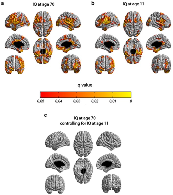
Associations between brain cortical tissue volume and cognitive function in old age are frequently interpreted as suggesting that preservation of cortical tissue is the foundation of successful cognitive aging. However, this association could also, in part, reflect a lifelong association between cognitive ability and cortical tissue. We analyzed data on 588 subjects from the Lothian Birth Cohort 1936 who had intelligence quotient (IQ) scores from the same cognitive test available at both 11 and 70 years of age as well as high-resolution brain magnetic resonance imaging data obtained at approximately 73 years of age. Cortical thickness was estimated at 81 924 sampling points across the cortex for each subject using an automated pipeline. Multiple regression was used to assess associations between cortical thickness and the IQ measures at 11 and 70 years. Childhood IQ accounted for more than two-third of the association between IQ at 70 years and cortical thickness measured at age 73 years. This warns against ascribing a causal interpretation to the association between cognitive ability and cortical tissue in old age based on assumptions about, and exclusive reference to, the aging process and any associated disease. Without early-life measures of cognitive ability, it would have been tempting to conclude that preservation of cortical thickness in old age is a foundation for successful cognitive aging when, instead, it is a lifelong association. This being said, results should not be construed as meaning that all studies on aging require direct measures of childhood IQ, but as suggesting that proxy measures of prior cognitive function can be useful to take into consideration.
大脑皮质组织体积与老年认知功能之间的关联经常被解释为,皮质组织的保护是成功认知老化的基础。然而,这种关联在某种程度上也可能反映了认知能力和皮质组织之间终生的关联。我们分析了来自洛锡安出生队列 1936 年的 588 名受试者的数据,这些受试者在 11 岁和 70 岁时都接受了相同认知测试的智商(IQ)评分,并且在大约 73 岁时还接受了高分辨率脑磁共振成像数据。使用自动流水线,为每个受试者的皮质表面的 81924 个采样点估计了皮质厚度。使用多元回归来评估皮质厚度与 11 岁和 70 岁时的 IQ 测量值之间的关联。儿童时期的智商解释了 70 岁时的智商与 73 岁时测量的皮质厚度之间 2/3 以上的关联。这告诫人们不要根据对衰老过程及任何相关疾病的假设和参考,将认知能力与老年皮质组织之间的关联归因于因果关系。如果没有儿童时期认知能力的测量,人们可能会轻易地得出结论,认为老年皮质厚度的保持是成功认知老化的基础,而实际上,这是一种终生的关联。话虽如此,这些结果不应被解释为意味着所有关于衰老的研究都需要直接测量儿童时期的智商,而只是表明先前认知功能的替代测量方法可以有用,值得考虑。